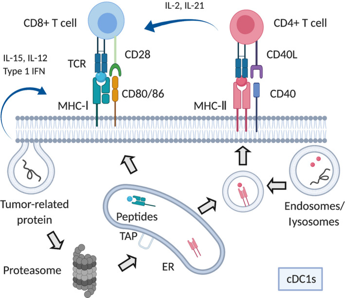
FIGURE 2.

Antigen presentation process. Tumor antigen‐derived proteins are taken up in antigen‐presenting cells by phagocytosis or endocytosis. In the MHC‐I pathway, proteins are degraded by the proteasome and enter into the endoplasmic reticulum (ER) through transporter associated with antigen processing (TAP). Subsequently, 8‐11 residues peptides are loaded onto the MHC‐I molecule and translocate to the cell surface where they could activate CD8+ T cells through cross‐presentation. In the MHC‐II pathway, endosomes or lysosomes take up tumor antigen‐derived proteins and digest them to 10‐30 (optimal 12‐16) residues peptides, which bind with MHC‐II for CD4+ T cell activation. Antigen‐specific CD4+/CD8+ T cells are activated by conventional type 1 dendritic cells (cDC1s) through T cell receptor (TCR) and costimulatory molecules. Various cytokines, including interleukin (IL)‐2 and IL‐21 from CD4+ T cells, especially type 1 helper cells, and IL‐15, IL‐12, and type I interferons from dendritic cells are produced. In part, these cytokines support differentiation and proliferation of CD8+ T cells
