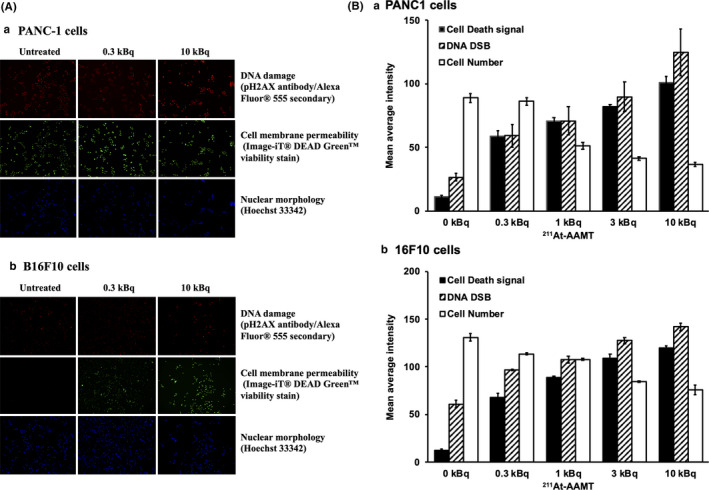
FIGURE 4.

Induction of DNA double‐stranded breaks by 211At‐AAMT in PANC‐1 cells. Imaging of 211At‐AAMT genotoxicity and cytotoxicity in PANC1 or B16F10 cells. Cells were treated with 0.3‐10 kBq of 211At‐AAMT for 5 min. Even at 0.3 kBq 211At‐AAMT, cells were positive for pH2AX, and the Image‐iT® DEAD GreenTM viability stain indicated DNA damage and compromise in plasma membrane integrity. Hoechst 33342 stain was used as a nuclear segmentation tool (A). The bar graph (B) shows the quantitative representation of 211At‐AAMT‐treated cells
