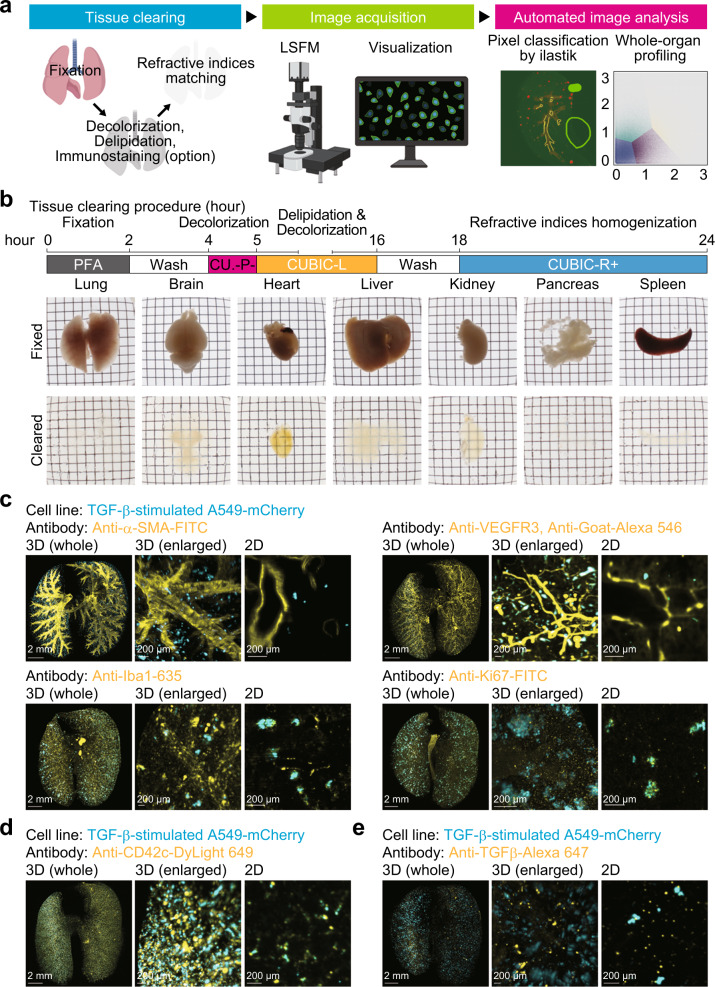
Fig. 1. Visualization of the tumour microenvironment using a one-day whole-organ clearing protocol.
a Scheme of whole-organ profiling of the tumour microenvironment using tissue clearing, image acquisition, and automated image analysis. LSFM light-sheet fluorescence microscope. b Protocol of one-day whole-organ clearing (top). Bright-field images of organs (lung, brain, heart, liver, kidney, pancreas, and spleen) after fixation (RI = 1.33) and clearing (RI = 1.52). Fixed organs were stocked in PBS buffer after PFA fixation. c Visualization of the tumour microenvironment in the experimental lung metastasis model. A549-mCherry cells were intravenously injected in mice (day 0). Then, the lung was subjected to whole-organ clearing protocol and immunostained with FITC-conjugated anti-α-SMA antibody (day 7), anti-VEGFR3 antibody, Alexa 546-conjugated anti-goat IgG antibody (day 1), Red Fluorochrome (635)-conjugated anti-Iba1 antibody (day 14), or FITC-conjugated anti-Ki67 antibody (day 14). d Visualization of the platelets in the experimental lung metastasis model. A549-mCherry cells were intravenously injected in mice (hour 0). Mice were administered with DyLight 649-conjugated anti-CD42c antibody immediately. Then, the lung was subjected to whole-organ clearing protocol, followed by 3D imaging (hour 1). e Visualization of TGF-β in the experimental lung metastasis model. A549-mCherry cells were intravenously injected in mice (day 0). Mice were administered with Alexa 647-conjugated anti-TGF-β antibody (day 6). Then, the lung was subjected to whole-organ clearing protocol, followed by 3D imaging (day 7). Representative images are shown. 3D image (whole), scale bar = 2000 μm. 3D image (enlarged), scale bar = 200 μm. 2D image, scale bar = 200 μm. Figure schematic created with biorender.com.

