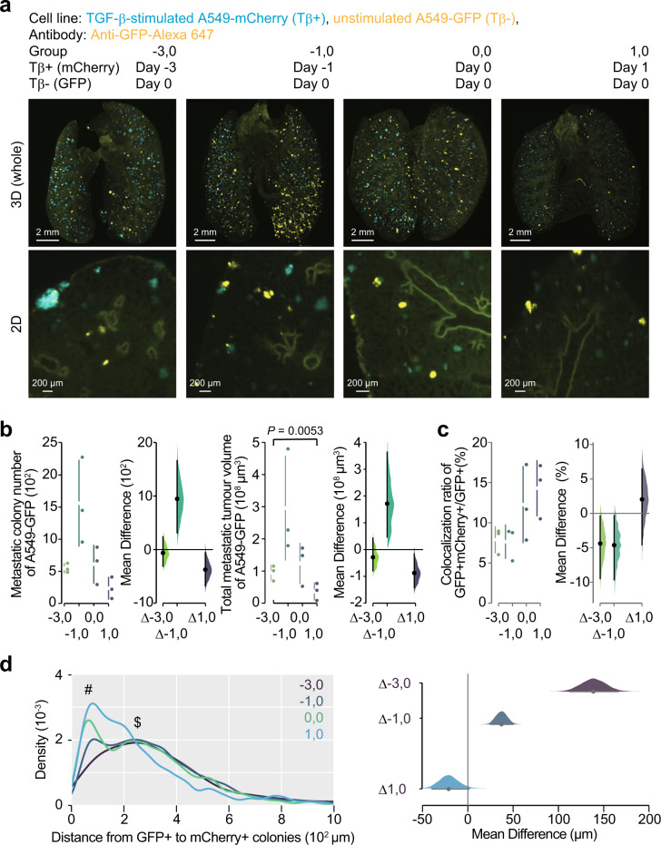
Fig. 4. TGF-β-stimulated cancer cells enhance colonization of unstimulated-cancer cells.
Temporal analysis of the effect of TGF-β-stimulated cancer cells on the metastasis of unstimulated cancer cells. Unstimulated A549-GFP cells were intravenously injected in mice (day 0). Same number of TGF-β-stimulated A549-mCherry cells were intravenously injected in the same mice at the indicated time point. Then, the lung was subjected to whole-organ clearing protocol (day 14), followed by 3D imaging. a Representative images. b Quantification of the metastatic colony number and the metastatic tumour volume of A549-GFP cells in lungs of mice. c Quantification of the colonies in which unstimulated cells were co-localized with TGF-β-stimulated cells. The ratios of GFP-positive and mCherry-positive colony number to the total GFP-positive colony number were indicated. d Density plot of the minimal distance from GFP-positive colony to mCherry-positive colony is shown. # and $ indicate the peaks around 50 µm and 250 µm, respectively. 3D image (whole), scale bar = 2000 μm. 2D image, scale bar = 200 μm. Mouse number in each group is n = 3. Representative result of two independent experiments. Data represent the effect size as a bootstrap 95% confidence interval.

