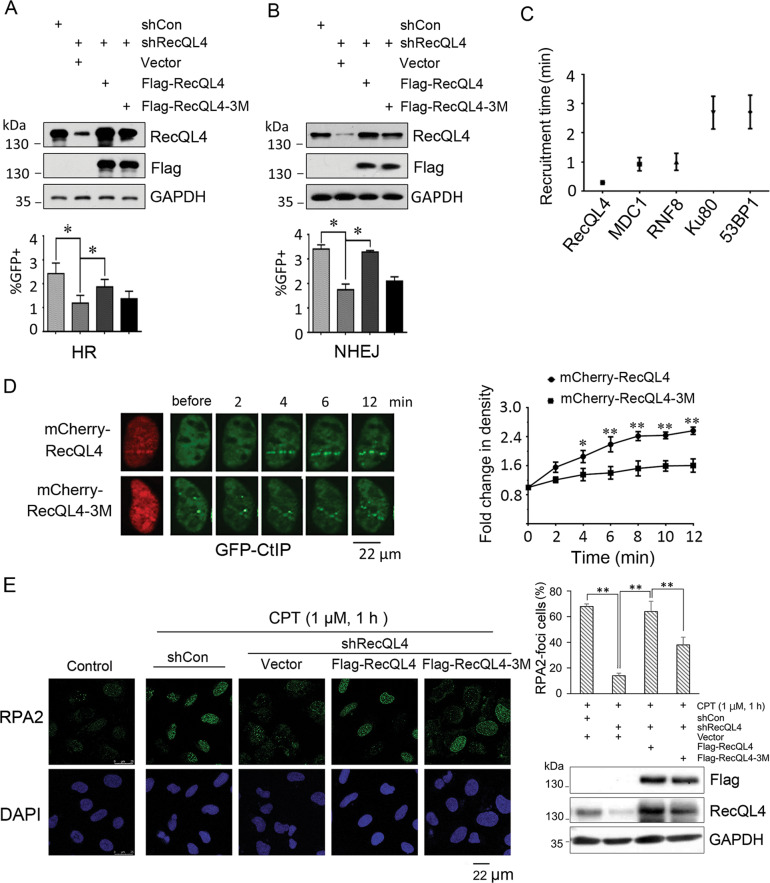
Fig. 4. Defective RecQL4 ubiquitination affects its activity in DSB repair and furthers the binding of its direct downstream proteins to DSB sites.
A, B RecQL4 depletion significantly decreased the HR- and NHEJ-mediated DSB repair in U2OS cells quantified by DR-GFP and EJ5-GFP reporter system, respectively. The defective DSB repair was significantly restored by re-introduction of wild-type RecQL4 but not its mutant. The percentage of GFP-positive cells was quantified by Flow cytometry. The data represent mean ± SEM from three independent experiments. (*p < 0.05, Student’s t test). C Analysis of the time-dependent recruitment of various DSB repair proteins in U2OS cells after micro-point laser treatment. Cells were transfected with GFP-tagged RecQL4, RNF8, Ku80, 53BP1, or mCherry-tagged MDC1 plasmids, followed by the treatment of micro-point laser. The images were captured using time-lapse microscopy. At least 15 cells for each transfection were recorded and analyzed for the earliest time point of protein aggregate formation at DSB track. D RecQL4 ubiquitination status affects the recruitment of its directly associated downstream protein-CtIP. RecQL4 was first silenced in U2OS cells followed by transfection with GFP-tagged CtIP and mCherry-tagged wild type RecQL4 or its mutant (3M). The mCherry-positive cells were treated with micro-point laser and the images were captured using microscopy. Both recruitment time and fluorescence density were recorded and at least 15 cells were analyzed. The data represent mean ± SEM from three independent experiments. **p < 0.01. E RecQL4 3M mutant interferes with its capacity in processing ssDNA formation at DSB ends estimated by an end resection assay. RecQL4 was first silenced by shRNA infection in U2OS cells which were then transfected with either a control, RecQL4 WT, or 3M mutant for 24 h, followed by the treatment with 1 μM CPT (C9911, Sigma) for 1 h. Cells were fixed for RPA2 (ab2175, Abcam) immunostaining. A total of 200 cells were analyzed and RPA2-foci (>15) positive cells were scored for each individual experiment. Data represent mean ± SEM of three independent experiments (**p < 0.01, the Single Factor Anova test). Fluorescence images were captured using a LEICA TCS SP8 confocal microscope system. Scale bar, 22 μm.

