
Figure 5.
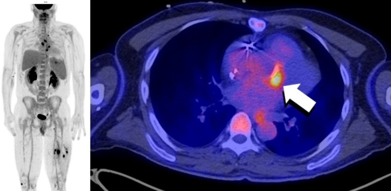
FDG PET/CT of a 51-year-old male status post aortic valve and ascending aorta replacement 10 years prior to the PET study. Multiple areas of abnormally increased FDG uptake are seen throughout on the body on the MIP images (left). Increased FDG uptake (arrow) is seen anteriorly to the aortic valve prosthesis (right). A focus of uptake in the spleen was compatible with a splenic abscess. Finally, areas of focally increased uptake are seen in both legs, with corresponding hypodensities on CT (not shown), compatible with intramuscular abscesses. The findings are compatible with an infectious endocarditis with septic emboli. Culture of the mechanical valve grew Staphylococcus capitis.