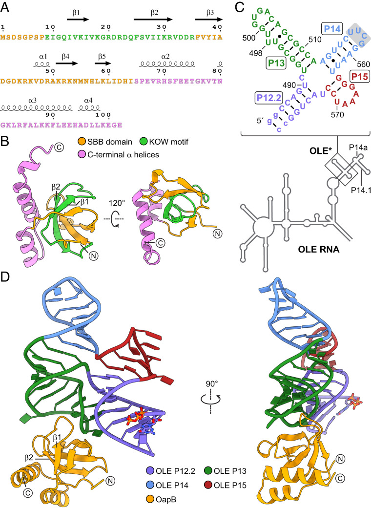
Fig. 1.
Constructs and structures of OapB and the OapB–OLE* complex. (A) Sequence and secondary structure and (B) crystal structure of B. halodurans OapB. The N-terminal SBB domain is colored orange, the C-terminal helices are colored violet, and the conserved 27-residue KOW motif is colored green. Encircled N and C letters denote the N and C termini of OapB. (C) Sequence and secondary structure model of the OLE* used in crystallization. Stem P14 is capped with a nonnative UUCG tetraloop (shaded box). Stem P12.2 is made more stable with two nonnative G-C base pairs (lowercase letters). (D) Front and side views of the OapB–OLE* complex crystal structure.

