Fig. 4.
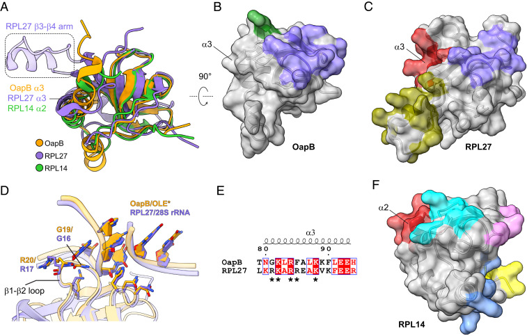
Comparison of OapB with KOW motif–containing ribosomal proteins. (A) Superimposition of OapB, human 60S ribosomal protein L27 (RPL27, PDB 6EK0) and human 60S ribosomal protein L14 (RPL14, PDB 6EK0) structures. (B) Surface representation of OapB. Separate RNA-interacting patches are colored, where purple represents the greatest similarity to the purple region in C. (C) Surface representation of RPL27. Separate RNA-interacting patches are colored. (D) Superimposition of the OapB–OLE* complex with RPL27-28S rRNA complex structures centered on their GNRA tetraloop regions reveals highly similar GNRA tetraloop–recognizing patterns by OapB and RPL27. Nucleotides are shown with filled rings. (E) Structure-based sequence alignment of α3 helices in OapB and RPL27. Amino acid residues in the basic patch (R108, K109, R111, R112, and K115) of RPL27 α3 are indicated with asterisks. The sequence alignment result was rendered using the online server of ESPript 3.0 (41). (F) Surface representations of RPL14.

