Fig. 3.
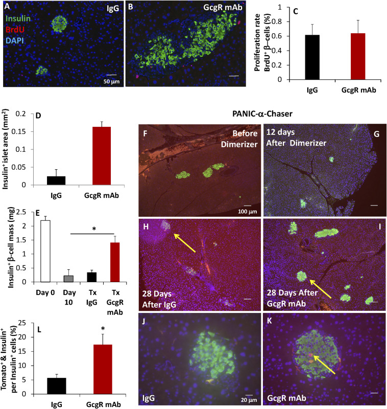
β-Cell regeneration and α-cell-to-β-cell conversion are increased after GcgR mAb treatment of PANIC-ATTAC mice. (A–C) BrdU was given 48 h prior to euthanasia, after 10 d of treatment with IgG (A) or GcgR mAb (B). n = 6 per group. (C) BrdU-positive β-cells were counted and expressed as percentage of insulin+ cells. (D) Average insulin+ pancreatic area was quantified. n = 18 per group. (E–K) td-Tomato expression was knocked-in to preproglucagon-expressing cells (using a doxycycline-inducible system) prior to the onset of diabetes. n = 8 per group. The formation of insulin+ (green) and td-Tomato+ (red) cells is seen in yellow, indicating insulin production from α-cell precursor cells. Insulin-positive islet mass was quantified from multiple nonadjacent sections following euthanasia. Yellow arrows indicate islets containing Tomato+ & Insulin+ cells. (L) Tomato+ & Insulin+ cells were counted and expressed as a percentage of Insulin+ cells. Standard error shown.

