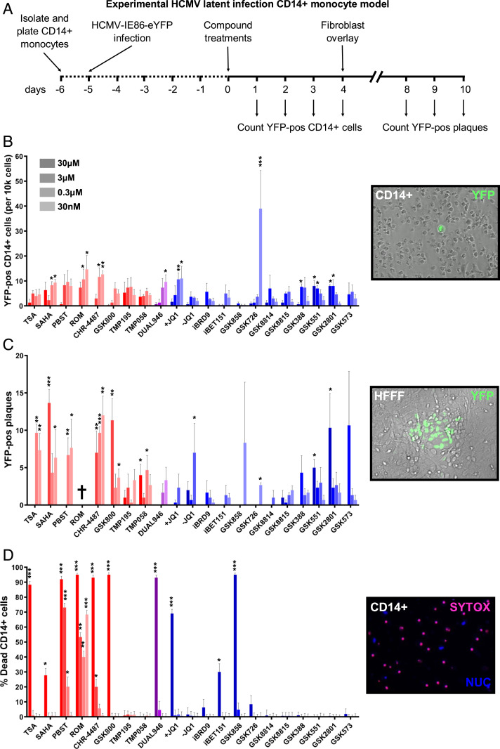
Fig. 2.
BET inhibitors induce virus gene expression while restricting full reactivation from ex vivo HCMV latency models. (A–C) CD14+ monocytes isolated from apheresis cones were infected with HCMV TB40e IE86-eYFP for 5 d before treatment with HDACi (red bars), BRDi/I-BET (blue bars), or a dual inhibitor (purple bars) across a range of concentrations (30 μM to 30 nM) for 72 h (A). The number of YFP-positive cells per well was then counted (B) before monocytes were overlaid with HFFFs and the resulting plaques were counted after 7 d (C) (mean + SEM, n = 3). (D) CD14+ monocytes isolated from apheresis cones were treated with inhibitors for 72 h before being stained with SYTOX (dead cells, red stain) and Hoechst stain (nucleus, blue stain) and counted (mean + SEM, n = 3) († = total cell death). (Insets) Example images of each condition counted (20× magnification). (*P < 0.05, **P < 0.01, ***P < 0.001.)

