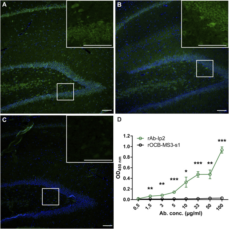
Fig. 1.
rAb-Ip2 recognizes the extracellular domain of GABAA-R-α1. Immunohistochemical staining of formalin-fixed paraffin-embedded rat hippocampus. (A) Staining with rAb-IP2 (green). Nuclei are stained with DAPI. Isotype control staining was negative. (Scale bar: 100 µm.) (B) Staining with the commercial antibody 62-3G1 to the GABAA-R-α1 subunit (green). (C) Staining with the negative control antibody rOCB-MS3-s1. (D) The ELISA shows that rAb-IP2 recognizes recombinant GABAA-R-α1ex produced in E. coli in a concentration-dependent manner (green). Control antibody rOCB-MS3-s1 does not show any reactivity to GABAA-R-α1ex (black). Error bars indicate SEM, n = 4. Statistical significance was calculated with GraphPad Prism 6 by unpaired t test. *P < 0.05,**P < 0.01,***P < 0.001.

