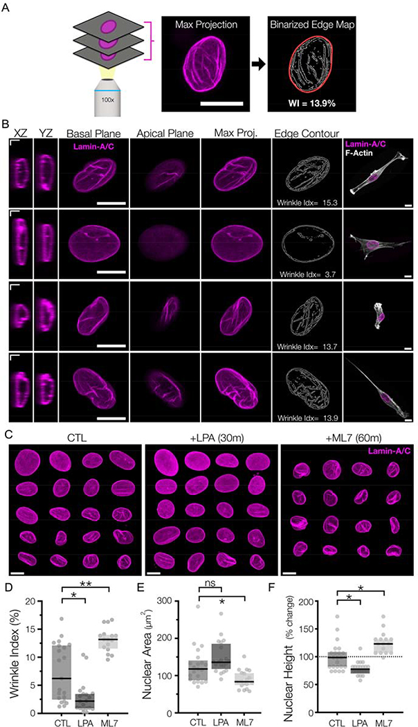
Figure #2-. Rapid alterations in NE morphology following pharmacologic changes in actomyosin contractility.
(A) Overview of the nuclear wrinkling metric based on edge detection. Maximum projection images of lamin-A/C z-stacks were taken and subjected to contrast-based Sobel edge detection. These resulting edge maps were binarized, and the fraction of wrinkled edges of these maps was normalized to the nuclear boundary area to generate the nuclear wrinkling index. SB = 5 μm. (B) Representative immunostaining of lamin-A/C from MSCs on 10 kPa hydrogels. Single confocal slices are shown of XZ/YZ slices (SB = 2 μm), as well as individual slices of the apical and basal sides of the nuclei. Maximum projection images are utilized to generate an edge-detection based contour map, which is used to generate a wrinkling index. SB = 10 μm. (C) Mosaic representations of Lamin-A/C max projection images for MSCs cultured on 10 kPa MeHA gels for 18 hours (“CTL”) or following treatment with LPA for 30m or ML7 for 60m at the end of this culture window. SB = 10 μm. Corresponding quantifications of (D) wrinkle index, (E) spread nuclear area, and (F) nuclear height of the nuclei from (C) (n>19 nuclei/group, Box plot represents median and 25th/75th quartiles, *= p<0.05, **= p<0.01, by Kruskal-Wallis one-way ANOVA testing with Dunn’s post-hoc).

