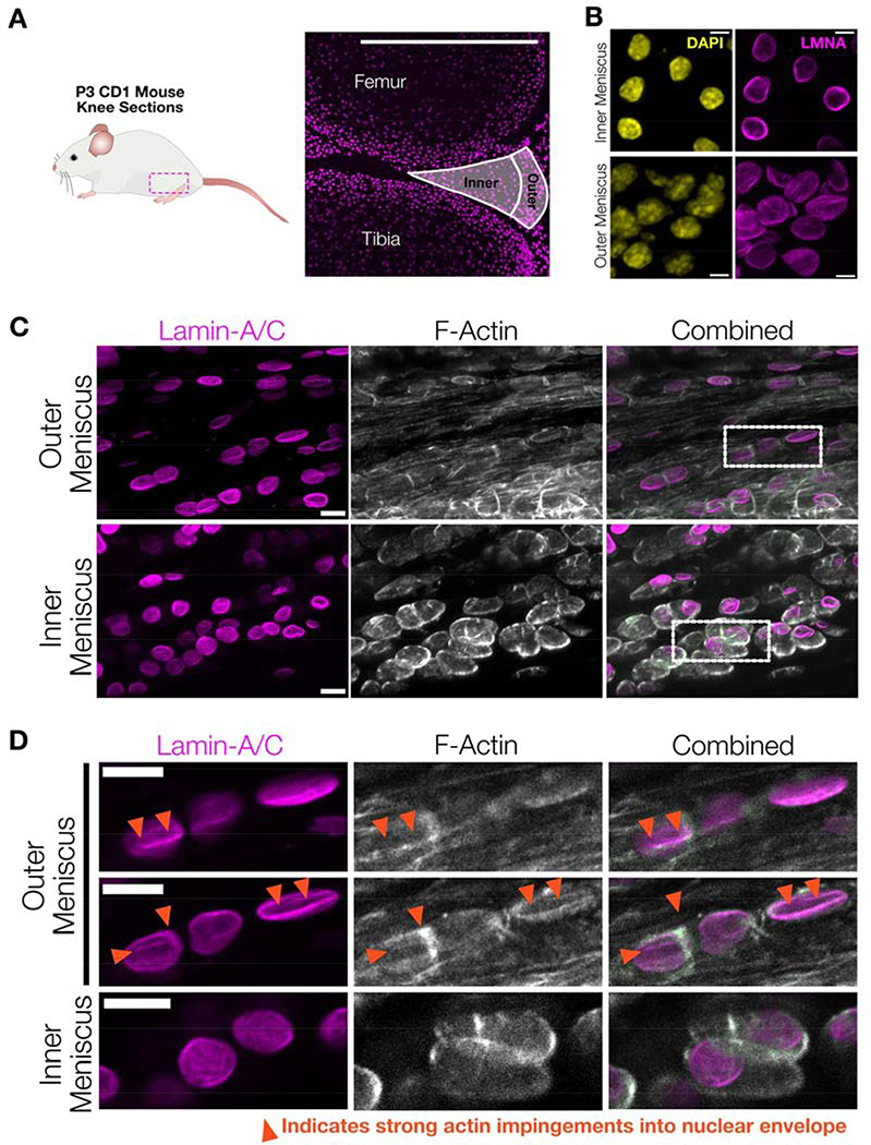
Figure #7 – Mesenchymal cells in juvenile mouse menisci have wrinkled nuclear envelopes with local actin impingement.
(A) To examine in vivo nuclear morphology of mesenchymal progenitor cells, 7.5 μm-thick sections were taken from the knee of P3 CD1 mice and immunostained. Cells from the inner and outer menisci were analyzed to characterize cell and nuclear morphology in the varying mechanical microenvironments present in the developing meniscus. SB = 500 μm. (B) Representative maximum projection z-stack images of chromatin (DAPI) and lamin-A/C across the inner and outer meniscus regions highlight increased nuclear envelope wrinkling in the outer meniscus region. SB = 5 μm. (C) Representative images taken from outer and inner meniscal regions highlighting fibrillar actin interactions with outer meniscus nuclei in P14 juvenile CD1 mice. SB = 10 μm. (D) Zoomed images from the dotted highlights in C, with two XY-sections from the same z-stack in the outer meniscus and one XY-section from the inner meniscus. These images highlight how fibrillar actin in mesenchymal progenitor cells residing in the outer meniscus can impinge on the nuclear envelope to create a wrinkled morphology (arrows), while mesenchymal progenitors residing in the inner meniscus exhibited more cortical actin structures and less wrinkled nuclei. SB = 10 μm.

