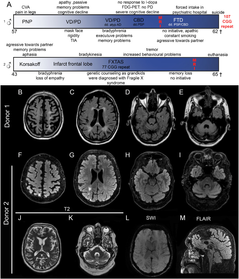
Figure 1.
Clinical course and imaging results of Donors 1 and 2. Clinical course (A) shows moment in time of MRI. General mild atrophy is observed (B, F). Ventricles are slightly dilated and Donor 1 shows periventricular white matter lesions (C), Donor 2 shows an increased signal due to an infarct (G). Slight cerebellar atrophy can be seen in both donors (D, E, H, I). Enlargement of the fourth ventricle, but no FXTAS-characteristic middle cerebral peduncle sign can be seen. T2-fluid-attenuated inversion recovery of Donor 2 does not show increased insular and middle cerebral peduncle signal intensities (J, K), no abnormalities in vasculature (K) and no thinning of the corpus callosum (M). CVA, cerebrovascular accident; PNP, polyneuropathy; VD, vascular dementia; PD, Parkinson’s disease; AD, Alzheimer’s disease; CBD, corticobasal degeneration; FTD, frontotemporal dementia; PSP, progressive supranuclear palsy.

