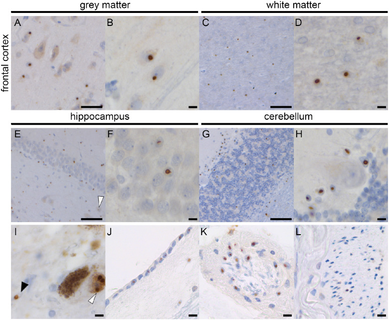
Figure 2.
p62-Pathology in Donor 2. Abundant p62-positive nuclear inclusions in the frontal cortex grey matter (A, B) and white matter (C, D). The hippocampus shows nuclear inclusions in the CA4 neuron (white arrowhead) and the dentate gyrus (E, F). In the cerebellum, inclusions are seen in the white matter and granular layer (G, H). No inclusions were seen in the Purkinje cells, whereas most Bergmann glia showed inclusions (H). Pathology was also seen in the dopaminergic neurons (white arrowhead) and astrocytes (black arrowhead) in the substantia nigra (I), ependymal (J), extra-axial cells of the third nerve (K) and choroid plexus cells (L). Scale bar A is 50 μm; Scale bar B, D, F, H, I–L is 10 μm; scale bar C, E, G is 100 μm.

