Figure 4.
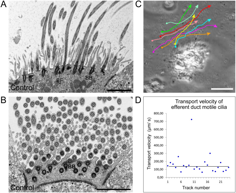
Mouse efferent duct cilia display a regular orientation and directed beating pattern. A and B, TEM analysis of ciliary cross sections in control mouse efferent ducts.A, longitudinal sections of efferent duct cilia displaying regular disposed basal bodies and basal foot processes. B, central pairs of efferent duct cilia (Ci) are oriented in a regular fashion as indicated by the red bars. C, manual tracking of 0.5 µm plastic beads applied on efferent duct cilia confirms that this cilia type displays a directed beating pattern. D, evaluation of the transport velocity of control efferent duct cilia shows that applied beads are mobilized with a median velocity of 130 µm/s. Biological replicate: n=3; Scale bars represent 2 µm (A and B), 50 µm (C).

