Figure 7.
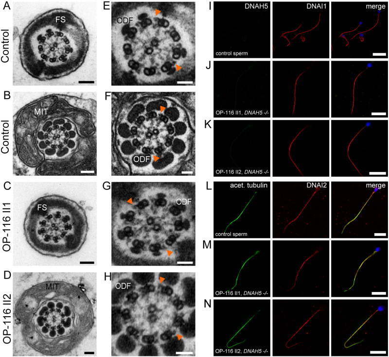
ODA protein DNAH5 is not present in human sperm flagella. A and B, TEM of control sperm cells showing the 9 + 2 axoneme with clearly visible ODAs and accessory structures (FS: fibrous sheath, MIT: mitochondria, and ODF: outer dense fibers). C and D, DNAH5 deficiency does not cause ODA defects of sperm flagellar axonemes. E–H, magnifications of A–D, orange arrowheads exemplarily indicate ODAs. I, J, K, IF co-staining assessing DNAH5 absence and panaxonemal localization of DNAI1 in human control and DNAH5-deficient sperm cells. L, control sperm cells display a panaxonemal flagellar localization of DNAI2 (red) that co-localizes (yellow in the merged image) with the flagellar marker acetylated alpha-tubulin (depicted in green). M and N, sperm flagella of individuals with PCD causing compound heterozygous mutations in DNAH5 display an unaltered localization pattern of DNAI2, indicating that DNAH5 is not a component of the ODA in sperm flagella. Nuclei (blue) were stained with DAPI. Scale bars represent 100 nm (A–D), 50 nm (E–H), 10 µm (I–N).

