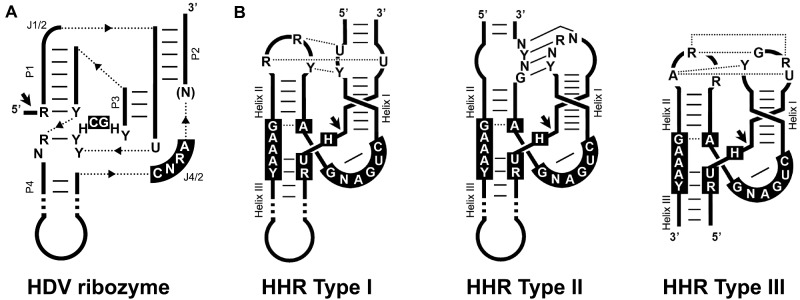
Figure 1.
(A) Schematic representation of the general topology of the hepatitis delta virus ribozyme (HDVR). Dotted lines represent connections between the helixes. The most frequent conserved nucleotides are shown in black boxes. Helical stems (P) and single-stranded junction strands (J) are indicated. (B) Schematic representation of the three possible hammerhead ribozyme (HHR) topologies (Types I, II, and III). The most frequent nucleotides in the catalytic core are shown in black boxes. Conserved loop-loop interactions are indicated (dotted and continuous lines refer to non-canonical and Watson–Crick base pairs). Black arrows indicate the self-cleavage site. The three HHR types have been reported in prokaryotic/phage genomes, whereas metazoan and plant genomes mostly show type I and III motifs, respectively.

