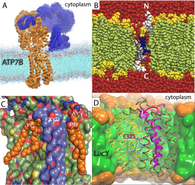
Fig. 1.
Membrane protein structural rearrangements and lipid regulation. a Homology model of the human Cu+-transporting P-type ATPase (ATP7B) (orange) with associated regulatory domain (blue) inserted into a lipid bilayer. Several simulated overlayed structures visualize thermal fluctuations displayed within a single state of the reaction. b A simulated S4 voltage-sensor peptide (silver with white GGPG flanks) showing bilayer distortion as the charged residues (blue) become solvated by lipid phosphates and water molecules (red). The lipid headgroups and tails are displayed in yellow and green, respectively. Adapted from Freites et al. (2005). c Lipid sites (A1 and B) hosting PC and PS lipids remodeled from a crystal structure at the cytoplasmic side of the Na+, K+ ATPase transporter. Adapted from Cornelius et al. (2015). d Lipid-dependent dynamics (magenta arrow and helix) and internal trigger (protonation state of residue E325) in the Lactose permease (LacY) transporter. The C-terminal and N-terminal domains are colored yellow and tan, respectively. The hydrophobic core of the membrane is colored green, and the polar headgroup region is depicted in orange. Adapted from (Andersson et al. 2012)

