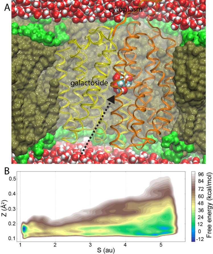
Fig. 3.
Sugar-binding energetics determined by metadynamics-enhanced sampling. a LacY crystal structure open towards the periplasm inserted into a lipid bilayer with hydrophobic core and polar headgroups in brown and green, respectively, with water molecules solvating both sides of the membrane. Extended, unbiased MD simulations identified a sugar-uptake pathway from the periplasm (dashed arrow). b Metadynamics simulations using the location of the uptake pathway (Z and S) as collective variables determined the free energy associated with sugar uptake and binding with excellent agreement to experimental data. Adapted from Kimanius et al. (2018)

