Fig. 1.

Example of meniscus segmentation
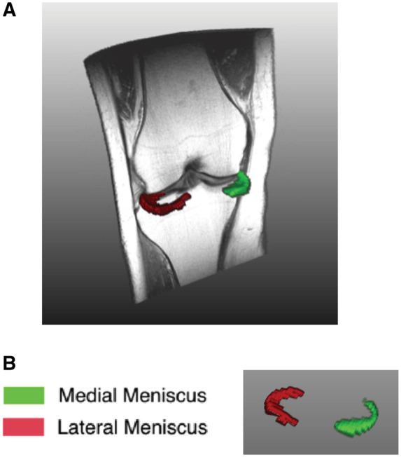
(A) 3D overview of one left knee and coronal view of meniscus segmentation. (B) 3D view of meniscus from segmentation (green: medial meniscus; red: lateral meniscus).

Example of meniscus segmentation
(A) 3D overview of one left knee and coronal view of meniscus segmentation. (B) 3D view of meniscus from segmentation (green: medial meniscus; red: lateral meniscus).