Fig. 3.
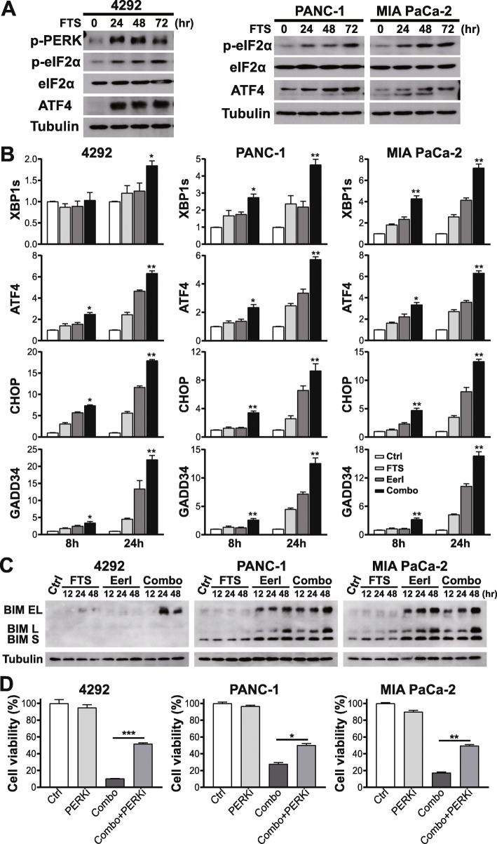
Activation of UPR by FTS is further enhanced upon ERAD inhibition to elicit ER stress-induced apoptosis. a Protein expression of UPR markers (p-PERK, p-eIF2α, ATF4) in PDAC cells treated with 100 μM FTS for 72 h. b Changes in transcript levels of UPR marker genes (XBP1s, ATF4, CHOP, and GADD34) following treatment with 100 μM FTS, EerI (3 μM for 4292 and MIA PaCa-2, 2 μM for PANC-1), or both for 8 and 24 h. All mRNA values were normalized to human GAPDH or mouse UBC. Two-tailed student’s t test compares transcriptional difference between combination and single treatments or control at the same timepoint, *P < 0.05, **P < 0.01; data presented as mean ± SD; n = 3. c Protein expression of BIM isoforms in PDAC cells in response to treatments with FTS, EerI, or the combination for 48 h. d Pretreatment with PERKi (2.5 μM GSK2606414) for two hours partially blocked the cell death induced by the combined FTS and EerI treatment for five days. Two-tailed student’s t test, *P < 0.05, **P < 0.01, ***P < 0.001; relative cell viability are presented as mean ± SD; n = 3

