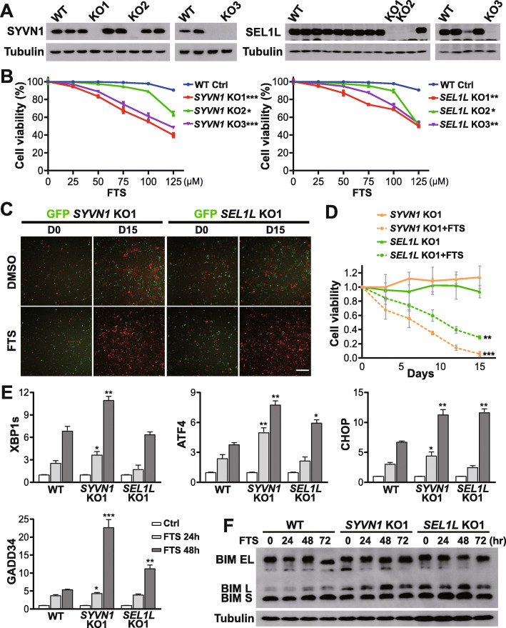
Fig. 4.
Knockout of SYVN1 and SEL1L sensitizes PDAC cells to FTS treatment through proapoptotic UPR. a Validation of SYVN1 and SEL1L CRISPR knockouts by western blot analysis of single clones derived from MIA PaCa-2 cells. b Relative cell viability of wild type control and SYVN1 and SEL1L KO clones treated with 25-125 μM FTS for five days. Two-tailed student’s t test, *P < 0.05, **P < 0.01, ***P < 0.001; data are presented as mean ± SD; n = 3. c Representative images of multicolor competition assay (50X) in the presence of 125 μM FTS. Scale bar, 100 μm. d Quantitative analysis of MCA for SYVN1 KO1 and SEL1L KO1 cell following FTS treatment. Unpaired two-sample t test, **P < 0.01, ***P < 0.001; data are presented as mean ± SD; n = 3. e Changes in transcript levels of UPR markers (XBP1s, ATF4, CHOP, and GADD34) in wild type control, SYVN1 KO1 and SEL1L KO1 cells following treatment with 100 μM FTS for 48 h. All mRNA values were normalized to human GAPDH. Two-tailed student’s t test compares transcriptional difference between KO clones and WT control at the same timepoint, *P < 0.05, **P < 0.01, ***P < 0.001; data are presented as mean ± SD; n = 3. (F) BIM protein expression in wild type control, SYVN1 KO1, and SEL1L KO1 cells following treatment with 100 μM FTS for 72 h

