Fig. 1.

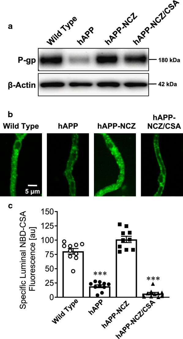
Effect of NCZ on P-gp protein expression and transport activity. a Western Blot for P-gp showing bands for brain capillary membranes isolated from vehicle-treated wild-type (WT), vehicle-treated hAPP mice, hAPP mice dosed with 5 mg/kg NCZ and hAPP mice dosed with a combination of NCZ and CSA (5/25 mg/kg). β-Actin was used as protein loading control; pooled tissue (WT: 10 mice; hAPP: 15 mice; hAPP-NCZ: 15 mice, hAPP-NCZ/CSA: 15 mice). Data were normalized to β-Actin. b Representative confocal images showing accumulation of P-gp-specific substrate NBD-CSA in isolated brain capillaries from WT, hAPP, hAPP-NCZ and hAPP-NCZ/CSA mice after a 1 hour incubation (steady state; 2 µM NBD-CSA). c Data after digital image analysis using ImageJ. Specific NBD-CSA fluorescence is the difference between total luminal NBD-CSA fluorescence and NBD-CSA fluorescence in the presence of the P-gp-specific inhibitor PSC833 (5 µM). Statistics: Data per group are given as mean ± S.E.M. for 10 capillaries from one preparation [pooled tissue: WT (10 mice), hAPP (15 mice), hAPP-NCZ (15 mice), hAPP-NCZ/CSA (15 mice)]. Shown are arbitrary fluorescence units (scale 0–255). ***, significantly lower than control, p < 0.001
