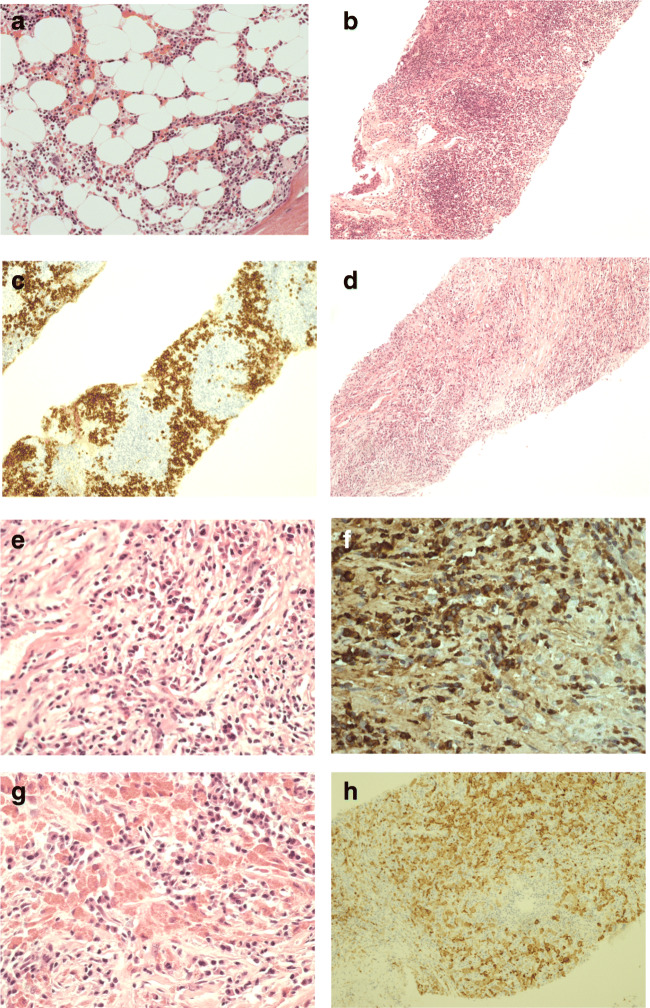
Fig. 1.
Histology of bone marrow, lymph node, and mediastinal mass biopsy. a Bone marrow trephine histology normal trilineage hematopoiesis, no plasma cell excess or macrophage infiltrate. b Lymph node biopsy showing retained architecture with B cell follicles and extensive plasmacytosis as shown in c lymph node biopsy CD138 IHC. Mediastinal mass biopsy (d) showed extensive fibrosis and mixed inflammatory infiltrate rich in plasma cells (e), with excess of IgG4 expressing plasma cells (f). In several areas, plasma cells were intermingled between macrophages with prominent eosinophilic cytoplasmic inclusions (g) and CD68 (h)

