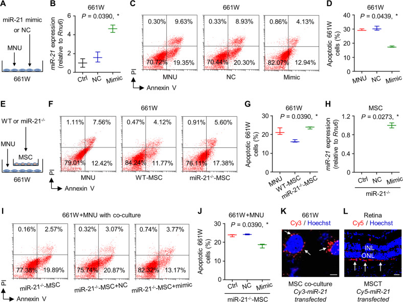
Fig. 6. miR-21 protects 661W cone photoreceptor cells against N-methyl-N-nitrosourea (MNU)-induced apoptosis and can be transferred from mesenchymal stem cells (MSCs).
A Diagram demonstrating the experimental design for analyzing direct effects of miR-21 on 661W cells. NC negative control of miR-21 mimics. B Quantitative real-time polymerase chain reaction (qRT-PCR) analysis of expression levels of miR-21 in 661W cells, normalized to Rnu6. Ctrl control. *P < 0.05 by the Kruskal–Wallis test. N = 3 per group. Representative flow cytometric images showing death of 661W cone photoreceptor cells (C) and the corresponding quantitative analysis of percentages of apoptotic (Annexin V+PI− plus Annexin V+PI+) 661W cells (D). 661W cells were treated with MNU with or without transfection of NC or miR-21 mimics. PI propidium iodide. *P < 0.05 by the Kruskal–Wallis test. N = 3 per group. E Diagram demonstrating the experimental design for analyzing indirect effects of MSCs on 661W cells. WT wild-type mice, miR-21−/−, mice deficient for miR-21. Representative flow cytometric images showing death of 661W cone photoreceptor cells (F) and the corresponding quantitative analysis of percentages of apoptotic (Annexin V+PI− plus Annexin V+PI+) 661W cells (G). MNU-treated 661W cells were co-cultured in the Transwell system with or without different MSCs. *P < 0.05 by the Kruskal–Wallis test. N = 3 per group. H qRT-PCR analysis of expression levels of miR-21 in MSCs, normalized to Rnu6. *P < 0.05 by the Kruskal–Wallis test. N = 3 per group. Representative flow cytometric images showing death of 661W cone photoreceptor cells (I) and the corresponding quantitative analysis of percentages of apoptotic (Annexin V+PI− plus Annexin V+PI+) 661W cells (J). 661W cells were treated with MNU in the Transwell co-culture system with MSCs, which were transfected with or without NC or miR-21 mimics. *P < 0.05 by the Kruskal–Wallis test. N = 3 per group. K Tracing of Cy3-labeled, MSC-derived miR-21 (red) in 661W cells during co-culture with MSCs, counterstained by Hoechst 33342 (blue). Scale bar = 10 μm. L Tracing of Cy5-labeled, MSC-derived miR-21 (red) in the retina tissue counterstained by Hoechst 33342 (blue) after intravitreal injection of MSCs for 24 h. MSCT mesenchymal stem cell transplantation, INL inner nuclear layer, ONL outer nuclear layer. Scale bar = 50 μm. Data represent median ± range for (B), (D), (G), (H), and (J).

