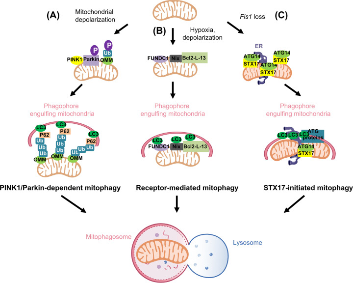
Fig. 2. Mammalian mitophagy machinery.
A During PINK1/Parkin-dependent mitophagy, ectopic toxins (such as CCCP) cause mitochondrial damage, in which PINK1 cleavage and import fail. PINK1 thus stabilizes on the outer mitochondrial membrane (OMM) and phosphorylates ubiquitin and Parkin. The attached OMM proteins are subsequently ubiquitinated by the E3 ligase, Parkin. These ubiquitinated OMM proteins serve as binding partners for P62. P62 further recruits LC3 and assembles the isolation membrane for selective autophagic mitochondrial removal. B During receptor-mediated mitophagy, in which cells are under hypoxic or mitochondrial membrane potential dissipation conditions, FUNDC1, Nix or Bcl-L-13 on OMM directly attracts LC3 via an LIR motif for mitophagosome formation. C STX17-induced mitophagy upon Fis1 loss participates in autonomous mitochondrial elimination through a hierarchical autophagic route. Under basal conditions, Fis1 interacts with STX17, preventing the over-translocation of STX17 onto mitochondria, governing the initiation of mitophagy. Fis1 loss primes the dynamic shuffling of STX17 onto mitochondria-associated membranes (MAM) and mitochondrial pools. Mitochondrial STX17 then self-oligomerizes and recruits ATG14. Subsequently, downstream autophagy modulators assemble on the mitochondria to ensure commitment to mitochondrial clearance.

