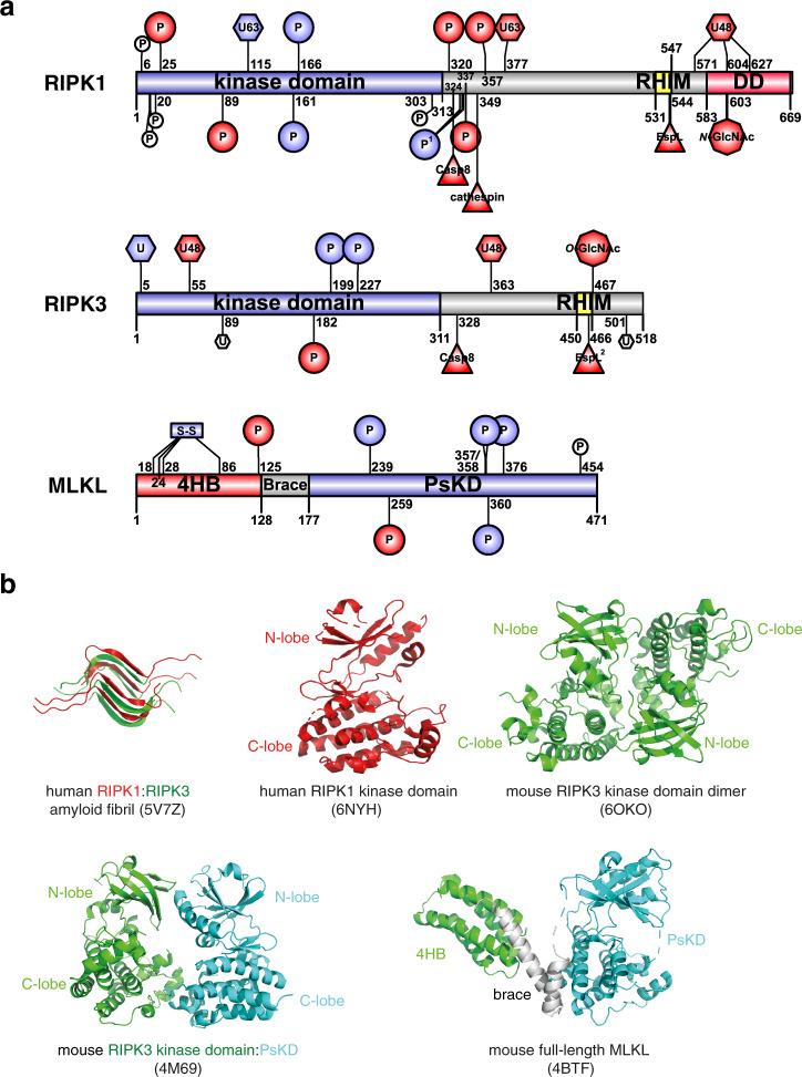
Fig. 4. Domain architecture and known post-translational modification sites of the necrosome components.
a Residues are numbered based on human ortholog sequence. Modifications are colored based on proposed effect to necroptotic cell death. Necroptosis-promoting (positive regulatory) events are colored blue; negative events are colored red. Modifications that were observed but have no known function in necroptosis are in white. Sequence diagrams were generated using IBS [204]. 4HB four-helix bundle domain, PsKD pseudokinase domain, RHIM RIP homotypic interaction motif, DD death domain. b Representative structures of RIPK1 and RIPK3 component domains and full-length MLKL (PDB accession numbers: 5V7Z [38]; 6NYH [71]; 6OZO [205]; 4M69 [105]; 4BTF [30]).

