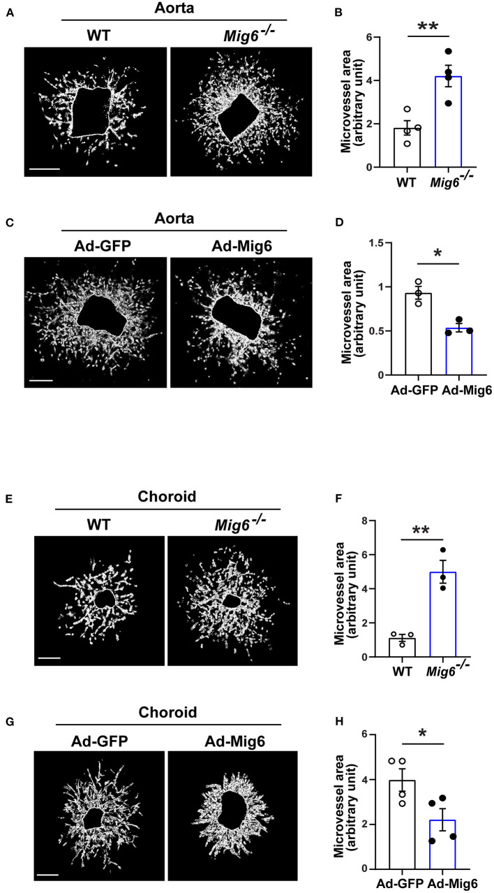
Figure 2.
MIG6 suppresses microvessel outgrowth. (A) Mouse aortic rings from WT or Mig6−/− aorta incubated for 7 days. (B) Quantification of vascular sprouting area per aortic ring in (A). n = 4 aortic rings per group. (C) Mouse aortic rings infected with Ad-GFP or Ad-MIG6 and incubated for 7 days. (D) Quantification of vascular sprouting area per aortic ring in (C). n = 3 aortic rings per group. (E) Mouse choroidal tissues of WT or Mig6−/− mice incubated for 7 days. (F) Quantification of vascular sprouting area per choroid tissue in (E). n = 3 choroids per group. (G) Mouse choroidal tissues infected with Ad-GFP or Ad-MIG6 and incubated for 7 days. (H) Quantification of vascular sprouting area per choroid tissue in (G). n = 3 choroids per group. Scale bars: 500 μm for (A); 250 μm for (C,E,G). The data are shown as the mean ± SEM from at least three independent experiments. * p < 0.05, ** p < 0.01 (two-tailed paired Student's t-test).

