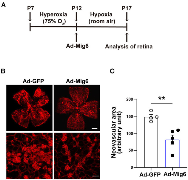
Figure 4.

MIG6 inhibits pathological angiogenesis. (A) Timeline of the retinopathy of prematurity model. P7 neonatal mice were exposed to hyperoxia for 5 days. Intravitreal injection of adenovirus expressing GFP or MIG6 was executed at P12. Mice were then returned to room air until P17. (B) Images of whole mount retinae at P17 stained with IB4-Alexa 594 (top). High magnification images of neovascular tufts in the retinae infected with Ad-GFP or Ad-MIG6 (bottom). (C) Quantification of neovascular areas in retinal whole mounts in (B). n = 4 fields per Ad-GFP treated retina, n = 5 fields per Ad-Mig6 treated retina. Scale bars: 300 μm for the top panel of (B); 50 μm for the bottom panel of (B). The data are shown as mean ± SEM. ** p < 0.01 (two-tailed paired Student's t-test).
