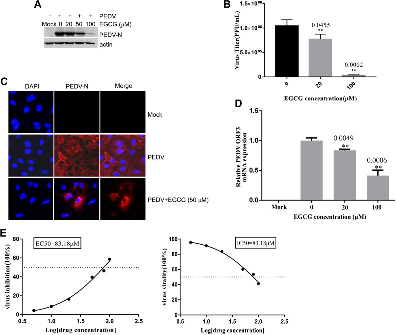
FIGURE 4.
EGCG inhibition of PEDV entry into Vero cells. (A–C) Vero cells were infected with PEDV HLJBY with EGCG present at 37°C for 1 h. After 1 h the cells were washed three times with citric acid and three times with PBS, and 2% DMEM was added. The culture was allowed to continue for 23 h at 37°C. (A) The level of PEDV N protein was evaluated by western blotting. (B) Plaque formation assay for PEDV titers in the supernatants. (C) The number of cells infected with PEDV was evaluated by IFA. (D) When the Vero cells were infected with PEDV with EGCG present at 37 C for 1 h, the PEDV ORF3 mRNA levels were detected by qRT-PCR. (E) EC50 or IC50 of EGCG against PEDV entry was checked.

