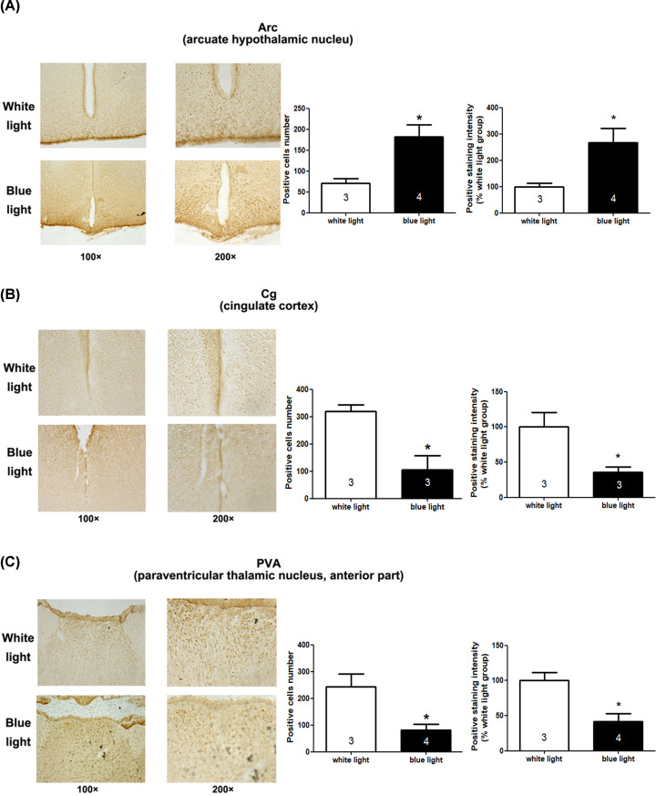
Figure 3. c-Fos expression in brain regions of mice exposed to white light or blue light.
c-Fos labelling of the arcuate hypothalamic nucleus (Arc) (A), cingulate cortex (Cg) (B) and anterior part of the paraventricular thalamic nucleus (PVA) (C). Positive cell numbers and positive staining intensity were analysed by unpaired t-test, showing that compared with the white light-treated mice, blue light induced significant changes in c-fos activation at Arc Cg and PVA (*P<0.05). The indicated number inside the column shows the number of mice calculated in each group.

