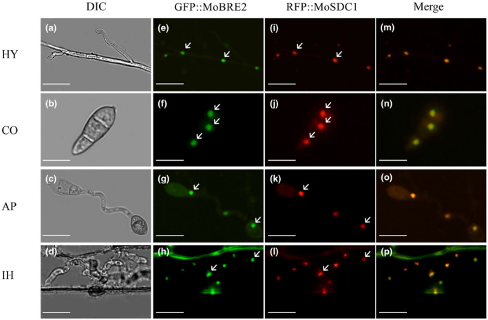
FIGURE 3.

Dual‐colour imaging of GFP‐MoBre2 and RFP‐MoSdc1 in Magnaporthe oryzae by confocal laser scanning microscopy. MoSdc1 was cloned into pKS, and the RFP‐MoSdc1 fusion construct was generated. The GFP‐MoBre2 and RFP‐MoSdc1 fusion constructs were digested with NotI and transformed into the protoplasts of wild‐type P131. The GFP‐MoBre2 and RFP‐MoSdc1 signals were merged. HY, vegetative hyphae; CO, conidia; AP, appressoria; IH, infection hyphae. DIC, differential interference contrast. Scale bars = 20 μm. Arrows indicate the nuclei localized in cells
