Figure 3.
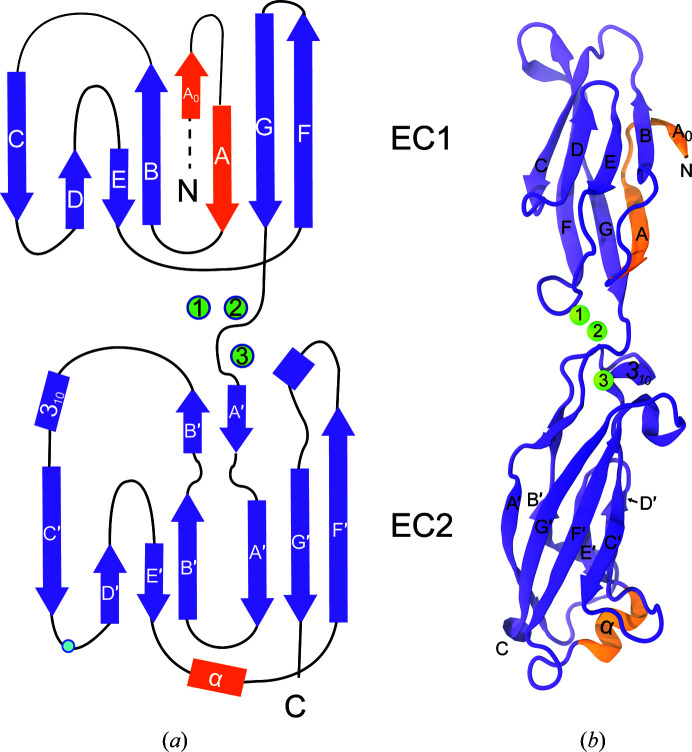
Structure of hs CDH17 EC1–2. (a) Topology of hs CDH17 EC1–2. Repeats EC1 and EC2 have canonical Greek-key motifs with atypical features in orange. The β-strands are labelled A–G for EC1 and A′–G′ for EC2, and α-helices are labelled by type. N and C denote the N- and C-terminus, respectively. Residue Asn154 is denoted by a blue dot. (b) Ribbon representation of the hs CDH17 EC1–2 structure with calcium ions in green and atypical features in orange. Labels are as in (a).

