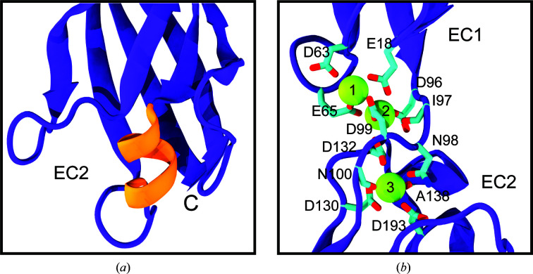
Figure 4.
Structural highlights of hs CDH17 EC1–2. (a) Detail of the two-turn α-helix in the EC2 E′–F′ loop, highlighted by the orange box in Fig. 1 ▸. (b) Detail of the hs CDH17 EC1–2 canonical calcium-binding linker. Side chains of calcium-coordinating residues are shown as sticks. Some backbone atoms are omitted for visualization purposes.

