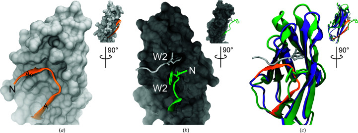
Figure 5.
Comparison of CDH17 and CDH1 EC1 repeats. (a) Detail of the hs CDH17 EC1 N-terminus showing residues Phe5–Glu18 in orange ribbon representation over the EC1 surface. (b) Detail of the hs CDH1 EC1 N-terminus (PDB entry 2o72; Parisini et al., 2007 ▸). The green strand shows EC1 residues Asp1–Glu11 extending out from the surface of the rest of EC1, while the silver strand shows the N-terminus of another monomer engaged in the strand-swap mechanism. A tryptophan residue (Trp2) is inserted in the hydrophobic pocket. (c) Rendering of hs CDH17 EC1 (purple and orange) structurally aligned with hs CDH1 EC1 (green) showing details of their contrasting N-termini. Insets in all panels show rotated views of EC1 highlighting the N-terminal strand location.

