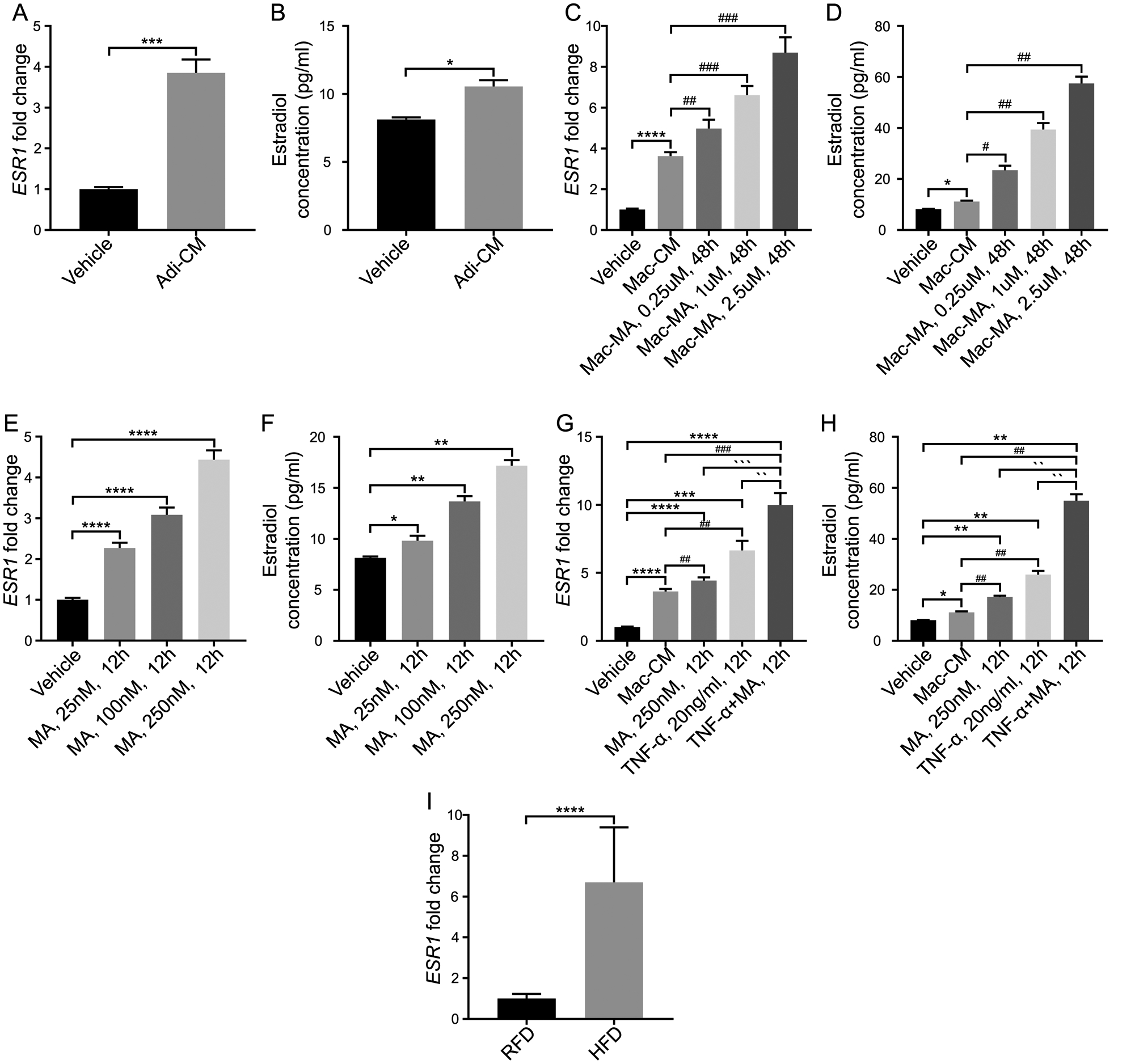
Fig. 4. Prostatic estrogen receptor-α (ESR1) and estradiol are elevated with adipocyte and inflammatory mediator exposure.

A–B: Stromal cells were treated with adipocyte conditioned media. ESR1 mRNA expressions and estradiol concentration were upregulated after treatment. C–D: Stromal cells were treated with macrophage conditioned media and activated-macrophage conditioned media. ESR1 mRNA expressions and estradiol concentration were upregulated in a dose-dependent manner after treatment. E–F: Stromal cells were treated with myristic acid. ESR1 mRNA expressions and estradiol concentration were upregulated in a dose-dependent manner after treatment. G–H: Stromal cells were treated with macrophage conditioned media (Mac-CM), MA, TNF-α and combination of MA and TNF-α. ESR1 mRNA expressions and estradiol concentration were upregulated after treatment of myristic acid, and the changes were more significant after treatment with TNF-α and combination of myristic acid and TNF- α. I: Prostatic ESR1 mRNA expressions and estradiol concentration were upregulated in mice with HFD exposure. *P < 0.05, **P< 0.01, ***P < 0.001, ****P< 0.0001, compared with RFD group in mice tissues, and compared with vehicle in stroma cells. ## P< 0.01, ### P < 0.001, compared with Mac-CM. `P < 0.05, ``P< 0.01, ```P < 0.001, ````P< 0.0001, compared with TNF-α +MA, 12h. Adi-CM: adipocyte conditioned media; Mac: macrophage conditioned media; MA: myristic acid.
