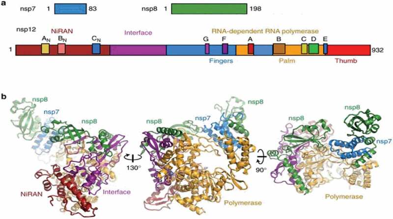
Figure 1.

Color-coded scheme and structure of the SARS-CoV nsp12 RdRp bound to nsp7 and nsp8 co-factors. (a) Diagram of the SARS-CoV nsp7, nsp8, and nsp12 proteins indicating domains and conserved motifs. (b) SARS-CoV nsp12 contains a large N-terminal extension composed of the NiRAN domain (dark red) and an interface domain (purple) adjacent to the polymerase domain (orange). nsp12 binds to a heterodimer of nsp7 (blue) and nsp8 (green) as well as to a second subunit of nsp8. Adapted (http://creativecommons.org/licenses/by/4.0/) from Kirchdoerfer et al. [12]. Color figure.
