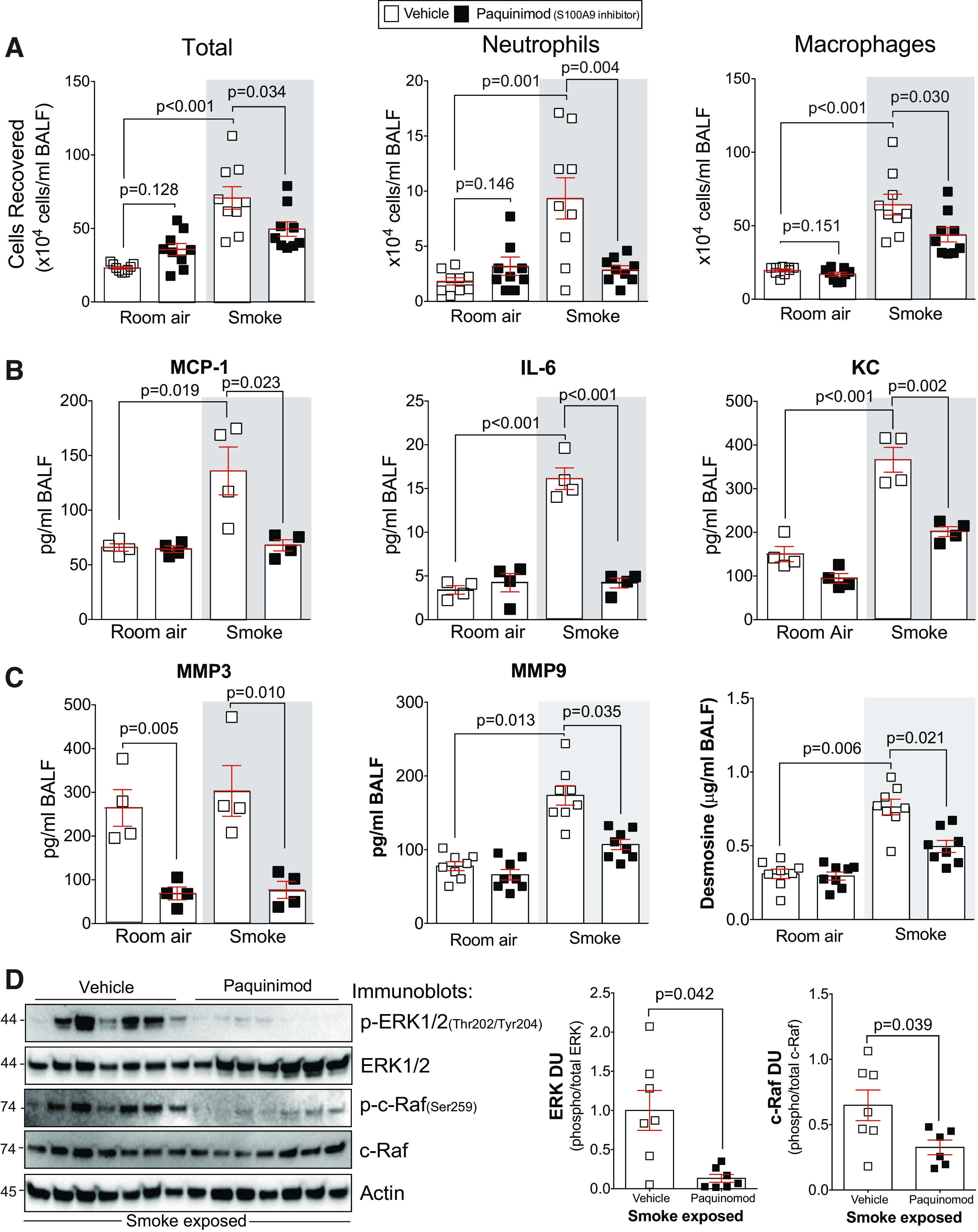
Fig. 6.

Paquinimod prevents inflammation and protease responses during smoke exposure. A: bronchoalveolar lavage fluid (BALF) cellularity levels were examined in A/J animals when paquinimod was administered daily before daily smoke exposure. BALF concentrations for MCP-1, IL-6, and keratinocyte-derived chemokine (KC) (B) and matrix metalloproteinase-3 (MMP-3), matrix metalloproteinase-9 (MMP-9), and desmosine (C) were quantified using Luminex bead assays. D: ERK and c-RAF phosphorylation were examined in lung tissue of smoke-exposed animals by Western blot and densitometry analysis. The representative blot shows data from seven animals per group. Graphs are represented as means ± SE, where n ≥ 5 per group. P values shown when comparing both treatments connected by a line were determined by two-way ANOVA with Tukey’s post hoc test. Graphs with two groups were analyzed by Student’s t tests.
