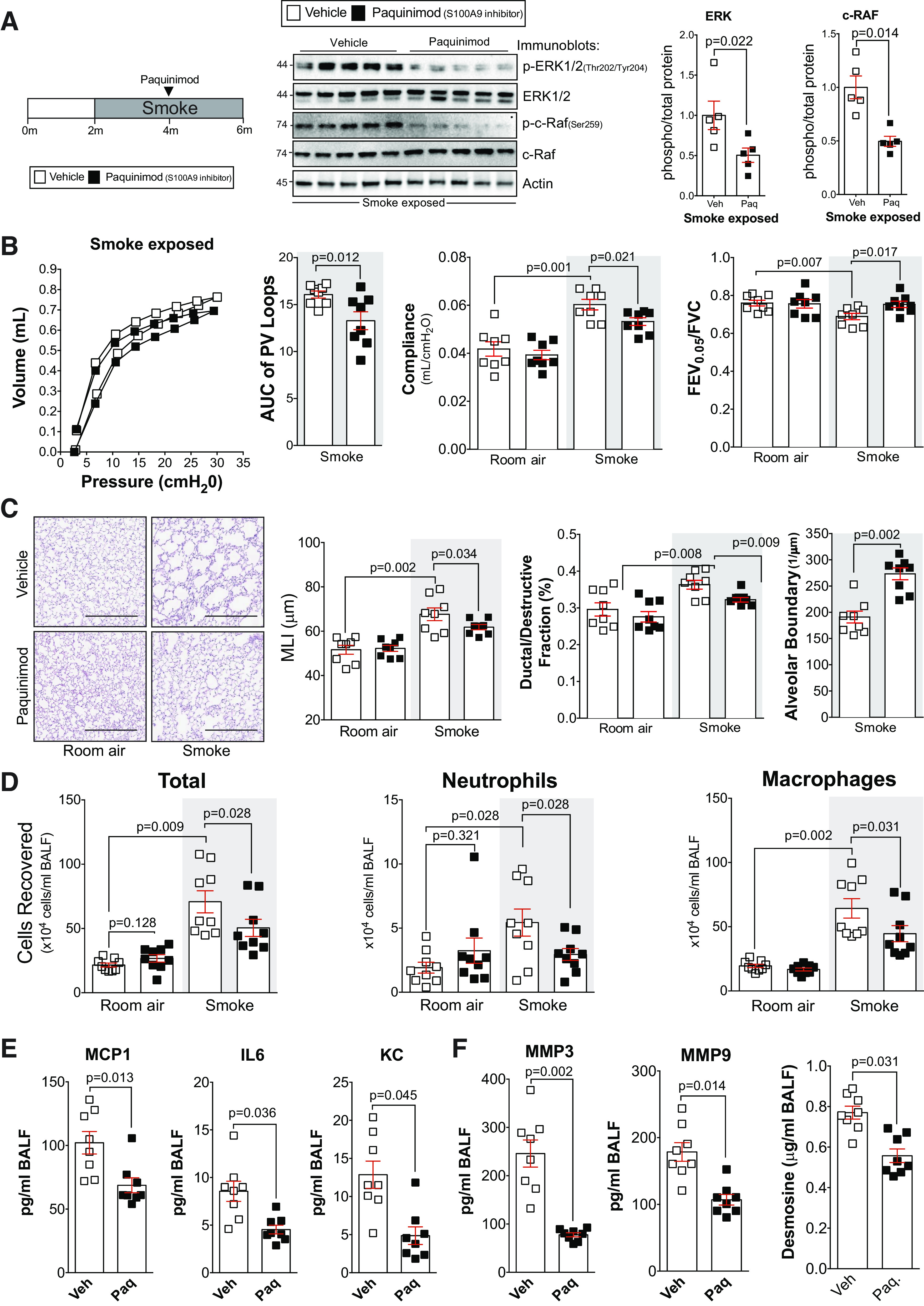
Fig. 7.

Delayed administration of paquinimod reduces the loss of lung function in smoke-exposed mice. A: A/J mice were smoke-exposed for 4 mo and began receiving paquinimod orally each day at the 2-mo mark after initiation of smoke exposure. ERK and c-RAF phosphorylation were examined in lung tissue of smoke-exposed animals by Western blot and densitometry analysis. B: forced expiratory volume (FEV) in the first 0.05 s of forced vital capacity (FVC), compliance, and pressure-volume loops with the area under the curve analysis were determined in each animal. C: mean linear intercept (MLI) was measured in the lungs of the mice to assess airspace size, and comparative histology images of the four mouse groups are presented here (scale bars = 500 µm). Alveolar count and ductal/destructive fractions were quantified in each animal by parenchymal airspace profiling. D: bronchoalveolar lavage fluid (BALF) cellularity levels were examined in A/J animals when paquinimod was administered daily before daily smoke exposure. BALF concentrations for monocyte chemoattractant protein-1 (MCP-1), IL-6, and keratinocyte-derived chemokine (KC) (E) and matrix metalloproteinase-3 (MMP-3), matrix metalloproteinase-9 (MMP-9), and desmosine (F) were quantified using Luminex bead assays. Graphs are represented as means ± SE, where n ≥ 5 per group. P values shown when comparing both treatments connected by a line were determined by two-way ANOVA with Tukey’s post hoc test. Graphs with two groups were analyzed by Student’s t tests.
