Fig. 1.
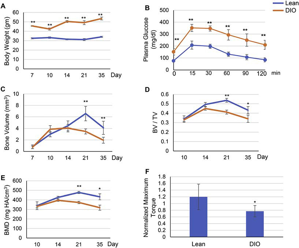
Fracture healing is delayed in DIO mice. (A) Body weight at time of harvest. Five-week-old mice were fed either lean or high-fat diet (denoted as Lean and DIO, respectively) for 12 weeks, subjected to mid-diaphysis tibial fracture, and harvested at the indicated post-fracture time points. (B) Pre-operative Glucose tolerance test (GTT). Mice were fasted for 6 h (daytime), received an IP injection of 2g/kg glucose, and blood glucose levels were measured at the specified post-injection time points. (C-E) Graphical presentation of μCT analysis of bone volume (C), bone volume density (BV/TV) (D), or bone mineral density (BMD) (E) performed at the specified post-fracture time points. (F) Graphical presentation of biomechanical torsional testing at day 35 post-fracture. Recorded maximum torque (N.mm) of the fractured right tibia was normalized to that of the contralateral unfractured left tibia to confirm that the observed decrease in biomechanical strength is specific to the healing tissue. For all analyses, n = 9. Data presented as average ± S.D. (*) P < 0.05; (**) P < 0.01 using student t-test and time-matched lean callus as a control.

