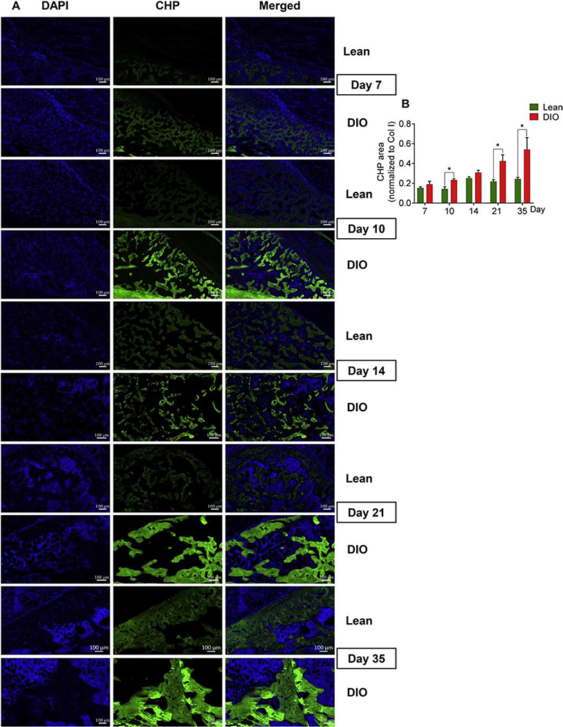
Fig. 7.
DIO callus has elevated levels of unfolded collagen helices. (A) Representative images of fluorescent staining (green) with collagen hybridizing peptide (CHP) that binds only to denatured, unfolded collagen strands. Staining was performed at the indicated post-fracture time points. Scale bar = 100μm. (B) Quantitation of CHP-stained area normalized to total Col I area.
Analysis was performed in 6 lean and 6 DIO callus tissues; 3 sections were analyzed in each. Bar graphs represent the average ± SEM. (*) P < 0.05 using student t-test and time-matched lean callus as a control.

