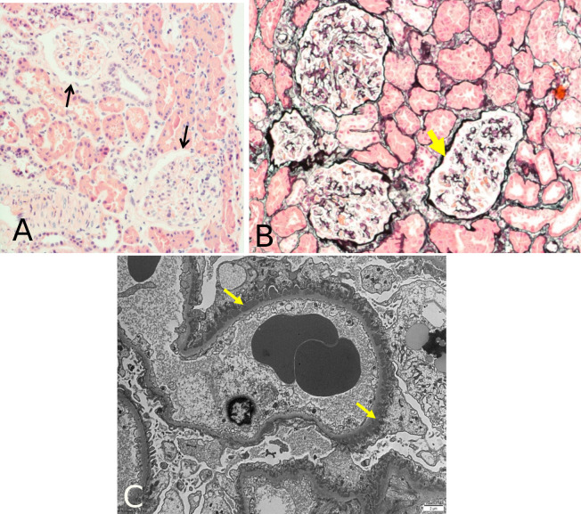
Figure 1.
Kidney biopsy results from light microscopy and electron microscopy. Light microscopy images did not actually showed a diagnosis of MN, with H&E stain (A) showing absence of thickening of GBM (arrows), and silver stain (B) showing absence of spikes along the GBM (arrow). When electron microscopy (C) was performed, small, electron dense, subepithelial deposits were observed with a strong diffuse granular IgG along the GBM (arrows), compatible with MN. GBM, glomerular basement membrane; MN, membranous nephropathy; H&E, Hematoxylin & Eosin.

