
Figure 2: Podocytes cultured on DCM display better viability.
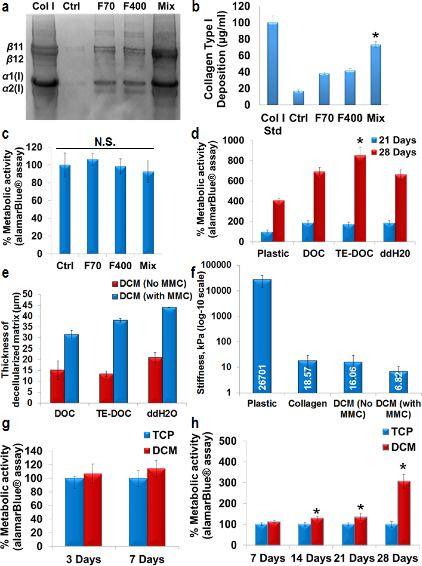
SDS-PAGE (a) and densitometry analysis (n=3) (b) of human skin fibroblasts cultured for 7 days. Maximum (*p<0.0001) collagen type I deposition was achieved under mix [Ficoll (F) 70 + F400] MMC conditions. Col I std – 100 μg/ml collagen type I from rat tail. alamar-Blue® analysis showed that there was no change (N.S. – no significance, p>0.05) in metabolic activity of fibroblasts cultured under MMC conditions (c). Significant increase (p<0.0001) in metabolic activity was observed in all the decellularization conditions in comparison with plastic for 21 days and 28 days. Decellularization solution with TE-DOC supported significantly higher metabolic activity (*p<0.0001, n=3) of podocytes up to 28 days (d). Different decellularization methods did not change the thickness of decellularized matrix. However, MMC significantly enhanced (p<0.0001, n=3) the thickness of decellularized matrix (e, thickness measured by SEM). AFM confirmed the higher stiffness of tissue culture plastic surface compared to collagen or DCM coated surfaces (f). Significant increase (*p<0.0001, n=3) in metabolic activity of podocytes was observed at various culture time (no difference in proliferation at 33° C, g) for differentiation at 37° C (h) and up to 28 days. All of the alamar-Blue® metabolic activity data were normalized for cell number. Ctrl – Control (no crowder added). SEM and AFM were done after 21 days of podocyte culture.