
Figure 3. Podocytes maturating on ECM produced by fibroblasts cultured in MMC conditions develop interdigitating foot processes.
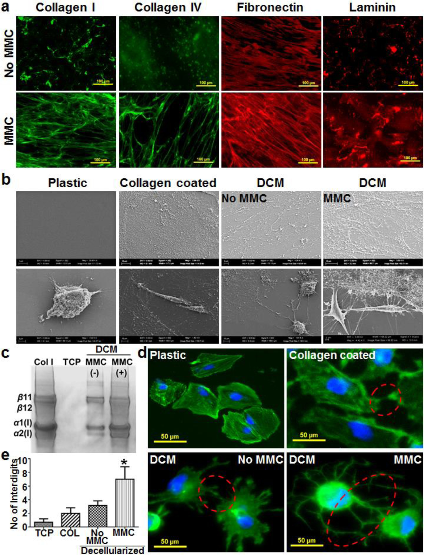
Immunofluorescence analysis of the tissue culture well plates with DCM confirmed higher deposition of collagen type I, IV, fibronectin and laminin under MMC conditions (a). Scanning electron microscopy of tissue culture plastic, collagen type I coated and cell derived decellularized matrix (DCM, with and without MMC) plates revealed the abundant deposition of fibrillar ECM in the DCM plates (b, upper panel) that helped podocytes to connect with matrix through their foot processes (b, lower panel). The collagen type I coated plastic plates did not show the presence of fibrillar matrix and assemblage between podocytes and matrix (b). Collagen type I was isolated using pepsin treatment and run on SDS-PAGE that further confirmed the higher deposition of collagen type I in DCM plates with MMC (c). Podocytes cultured (21 days) in DCM plates developed interdigitating foot processes among neighboring cells (d) and the number of interdigits between podocytes was significantly higher (*p<0.0001, n=15) in DCM plates with MMC (e), 15 images (3 images from 5 different culture wells were analyzed to count interdigits. TCP – Tissue culture plastic plates. DCM – Cell derived decellularized matrix well plates. COL – Tissue culture plastic plates coated with collagen type I. Col I std – 100 μg/ml collagen type I from rat tail.