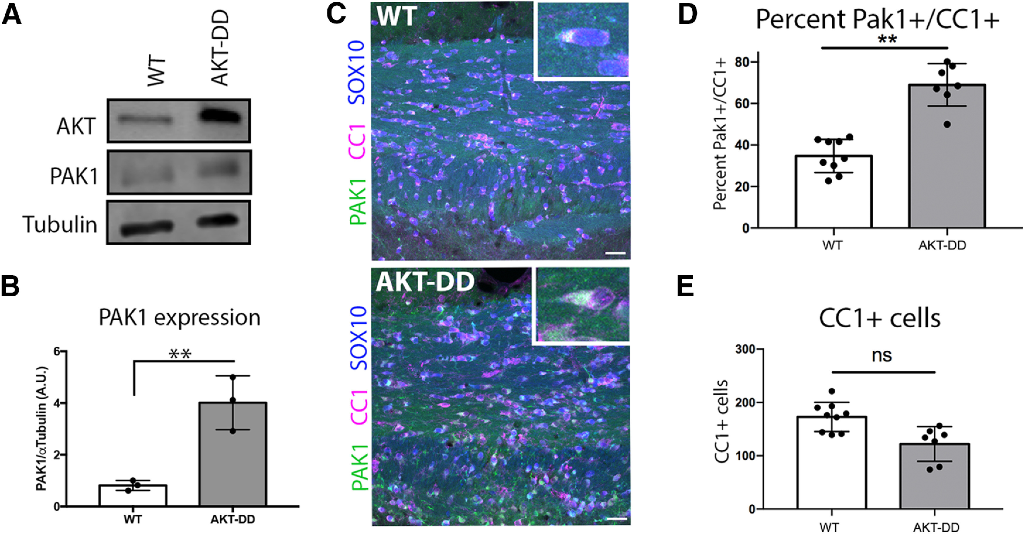
Figure 2.

Constitutively active AKT increases PAK1 protein expression in oligodendrocytes. A, Representative image of Western blot of WT or AKT-DD cerebellar protein samples, where AKT is overexpressed in PLP-Akt1 mice. B, Quantification of PAK1 protein expression compared with tubulin in WT and AKT-DD cerebellum samples of adult mice. n = 3 animals/genotype. t = 5.225, df = 4. **p = 0.0064 (unpaired t test). C, PAK1 is increased in oligodendrocytes in AKT-DD adult mice in the corpus callosum. Green represents PAK1. Blue represents SOX10 (entire oligodendrocyte lineage). Red represents CC1 (differentiated oligodendrocytes). Scale bar, 25 µm. D, E, Quantification of the percent of PAK1+/CC1+ cells and of total CC1+ cells. n = 3 animal/genotype, 2 or 3 images per animal. t = 8.467, df = 4. **p = 0.0011.
