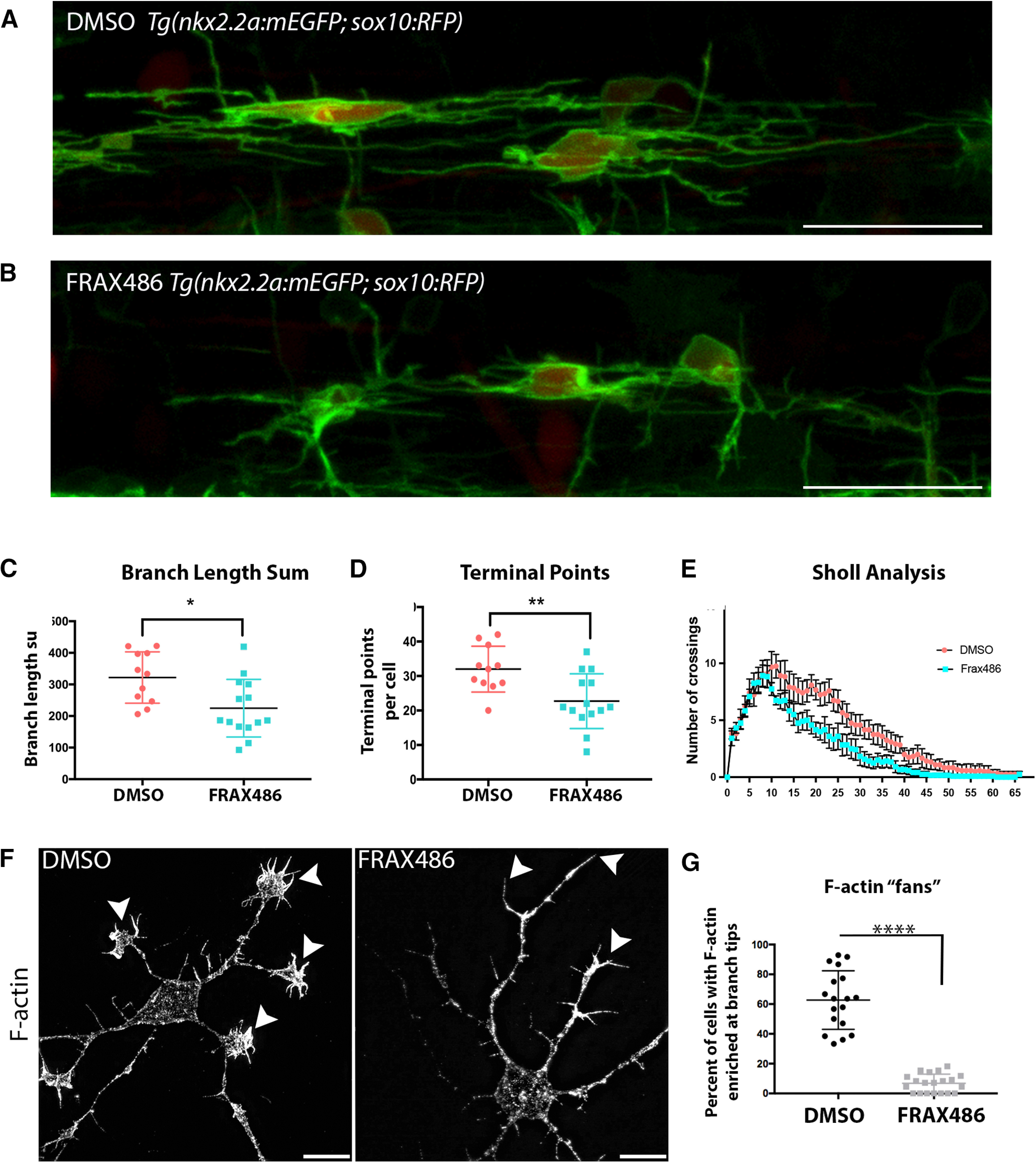
Figure 4.

Pharmacologically inhibiting PAK1 activity decreases oligodendrocyte branching complexity and reduces actin in OPC branches. A, B, Tg(nkx2.2:mEGFP; sox10:tagRFP) embryos were treated with a DMSO vehicle control (A) or the PAK1 inhibitor FRAX486 (B) from 48 to 56 hpf. Images were taken directly above the yolk sac extension. Individual cells were traced and analyzed in 3D using IMARIS software. Scale bar, 25 µm. C, The inhibition of PAK1 decreased the total length of oligodendrocyte branches. t = 2.771, df = 23. *p = 0.01 (unpaired t test). D, The inhibition of PAK1 decreased the number of oligodendrocyte terminal points per cell. t = 3.11, df = 23. **p = 0.0049 (unpaired t test). E, Sholl analysis was completed using IMARIS software. F, Mouse OPCs were treated for 30 min with DMSO vehicle control or the PAK inhibitor FRAX486 after 2 d in OPC media. Cells were fixed and stained for F-actin with phalloidin. Representative images at 100× are shown. Scale bar, 10 µm. Arrowheads point to the ends of OPC processes. There is dramatic loss of fan-like protrusions after FRAX486 treatment. N = 3 biological replicates with 5-7 technical replicates per group. G, Quantification of the percent of cells with enriched F-actin in fan-like protrusions at the ends of cell processes (phalloidin stain). Images were taken at 63×, and the number of Olig2+ cells with F-actin enriched in fan-like protrusions at the ends of cell processes was quantified. White represents DMSO. Gray represents FRAX486-treated cells. t = 12.09, df = 36. ****p < 0.0001 (unpaired t test).
