Figure 3.

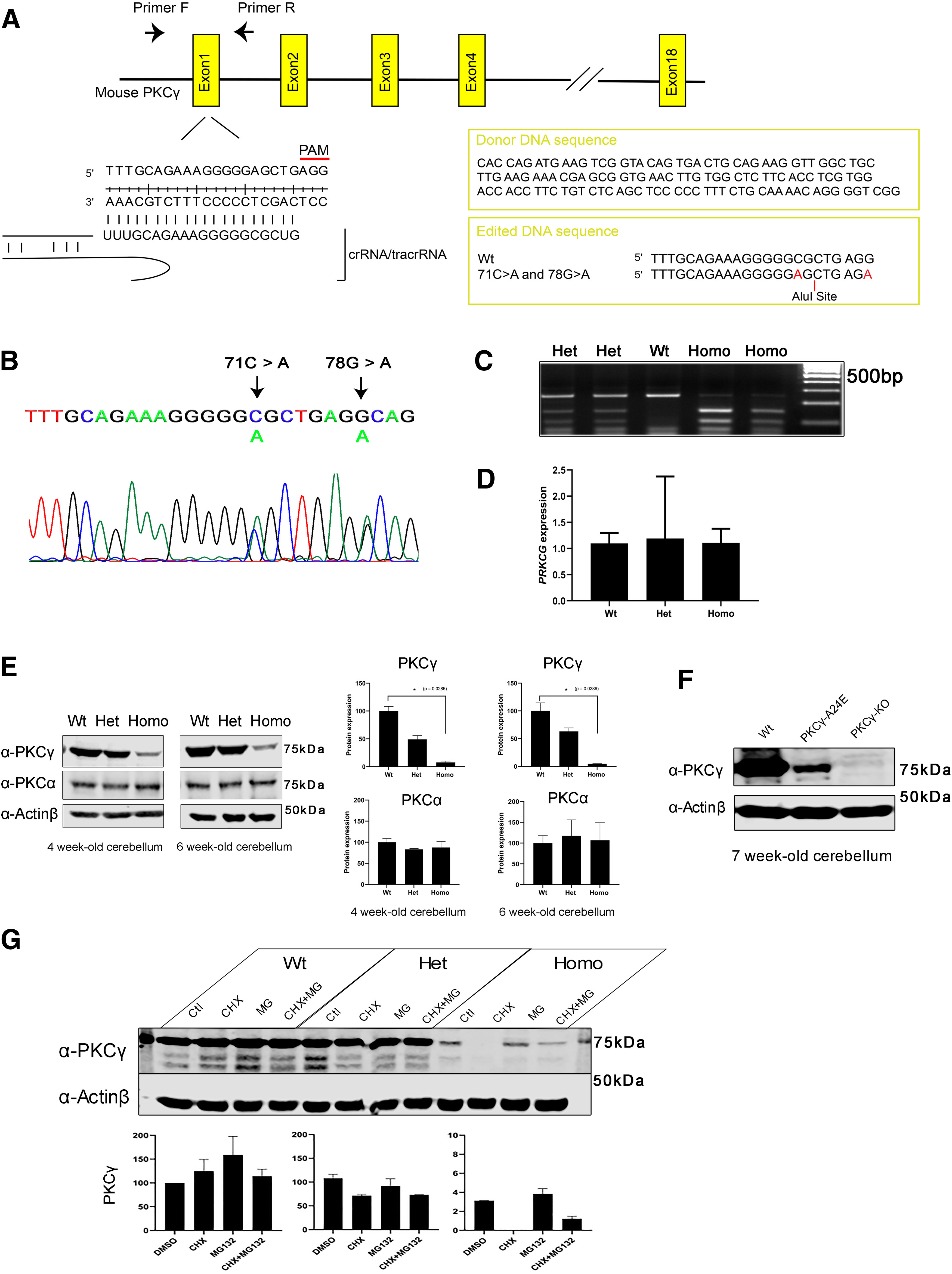
Generation and characterization of the PKCγ-A24E mouse. A, Target sequence in the PRKCG gene (chromosome 7, 1.93 cM) for producing point mutated (c. 71C > A; p. Ala 24 Glu) PRKCG with Cas9/CRISPR engineering system. To prevent donor DNA cleavage, the PAM sequence mutation was also introduced (78G > A), which does not give a change in amino acids. B, Mutations were validated with sequencing. The figure shows Het PKCγ-A24E sequence. C, To identify the genotypes, PCR was performed followed by Alu1 (BioLabs, R0137S) digestion for 1 h, which digests only Ala 24 Glu mutated DNA. Wt shows a single band (250 bp), Het PKCγ-A24E shows a Wt band and two digested bands (250, 150, and 100 bp), and Homo PKCγ-A24E shows only two digested bands (150 and 100 bp). D, qPCR using 5-week-old cerebellar samples from each genotype. Data show that all three genotypes express PKCγ mRNA. E, Western blot analysis of total PKCγ protein in the cerebellum from the three genotypes (n = 4). PKCγ protein degradation in PKCγ-A24E mice was observed (4-week-old: Wt = 100.0 ± 8.34%, n = 4; Het = 49.12 ± 6.80%, n = 4; Homo = 7.519 ± 2.48%, p = 0.0286, n = 4; 6-week-old: Wt = 100.0 ± 14.56%, n = 4; Het = 63.09 ± 6.39%, n = 4; Homo = 4.674 ± 0.40%, p = 0.0286, n = 4), whereas PKCα protein expression did not change in all genotypes (4-week-old: Wt = 100.0 ± 9.30%, n = 4; Het = 83.13 ± 2.09%, n = 4; Homo = 87.67 ± 14.1%, n = 4; 6-week-old: Wt = 100.0 ± 8.94%, n = 4; Het = 117.4 ± 19.3%, n = 4; Homo = 106.8 ± 21.1%, n = 4). The total expression levels of PKCγ and PKCα were normalized to the Actinβ expression level, and the expression levels of PKCγ and PKCα in Wt are shown as 100%. Data are mean ± SEM (n = 4, the statistical analysis showed no significance). Differences between Wt and Homo PKCγ-A24E were analyzed using the two-tailed Mann–Whitney test. F, Western blot analysis of total PKCγ protein in cerebellum from 7-week-old Wt, PKCγ-A24E, and PKCγ KO mice. PKCγ protein expression in Homo PKCγ-A24E mouse is confirmed via Western blot with long exposure, whereas PKCγ KO mouse shows no PKCγ protein expression (Wt = 100.0%, n = 4; Homo PKCγ-A24E = 2.088%, n = 4; PKCγ KO = 0.067%, n = 4). G, Organotypic slice culture from Wt, Het, and Homo PKCγ-A24E mice treated with 50 µg/ml cycloheximide or 10 μm MG132 or both at DIV7; protein was extracted at DIV8. Western blot data show that PKCγ protein expression is strongly reduced in PKCγ-A24E mice after cycloheximide treatment, whereas MG132 treatment rescued protein degradation in PKCγ-A24E mice. The total expression level of PKCγ was normalized to Actinβ expression level and DMSO-treated Wt is shown as 100.0% (Wt + DMSO = 100.0%; Wt + CHX = 124.9%; Wt + MG132 = 159.0%; Wt + MG132 + CHX = 114.2%; Het + DMSO = 107.8%; Het + CHX = 71.3%; Het + MG132 = 91.7%; Het + MG132 + CHX = 73.0%; Homo + DMSO = 3.1%; Homo + CHX = 0.03%; Homo + MG132 = 3.8%; Homo + MG132 + CHX = 1.2%, n = 2).
