Figure 6.
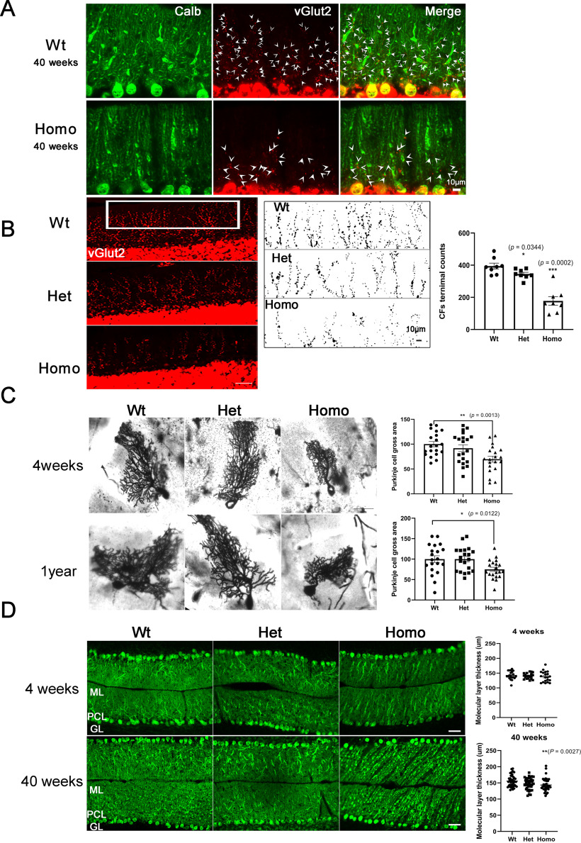
Altered CF innervation and Purkinje cell morphology in vivo in PKCγ-A24E mice. A, Anti-Calbindin D28K antibody staining shows Purkinje cells, and anti-vGlut2 staining shows CF terminals on Purkinje cell dendrites from 40-week-old Wt and Homo PKCγ-A24E mice. White arrowheads indicate CF terminals. Scale bar, 10 µm. B, The CF terminals on Purkinje cells were analyzed with anti-vGlut2 immunoreactivity. Scale bar, 50 µm. The CF terminals in the molecular layer were counted by ImageJ. Quantification of the number of terminal puncta on Purkinje cell branchlets in distal molecular layer in a 284 μm × 75 μm rectangle (vGlut2-positive puncta Wt = 394.4 ± 18.14; Het = 345.1 ± 10.61, p = 0.0344; Homo = 177.6 ± 26.62, p = 0.0002). Data are mean ± SEM of 8 samples each. Scale bar, 10 µm. C, Bright field microscopy analysis of Golgi-Cox staining from 4-week-old and 1-year-old Wt, Het, and Homo PKCγ-A24E mice shows significantly reduced size of Purkinje cells in Homo PKCγ-A24E mice. Purkinje cell size from 4-week-old (Wt = 100.0 ± 5.36%; Het PKCγ-A24E = 91.85 ± 6.81%; Homo PKCγ-A24E = 69.74 ± 6.07%) and Purkinje cell size from 1-year-old (Wt = 100.0 ± 8.17%; Het PKCγ-A24E = 99.85 ± 6.51%; Homo PKCγ-A24E = 74.91 ± 5.14%) were analyzed using the two-tailed Mann–Whitney test (4-week-old, p = 0.0013, 1-year-old, p = 0.0122). Scale bar, 50 µm. The number of measured cells was 20 for all experiments. D, Calbindin D-28 immunohistochemistry staining of cerebellar cryosections (20 µm) from 4-week-old to 40-week-old Wt, Het PKCγ-A24E, and Homo PKCγ-A24E mice. ML, Molecular layer; PCL, Purkinje cell layer; GL, granule cell layer. Scale bar, 50 µm. The width of the molecular layer is slightly decreased in lobule VII of 40-week-old Homo PKCγ-A24E mice (p = 0.0027).

