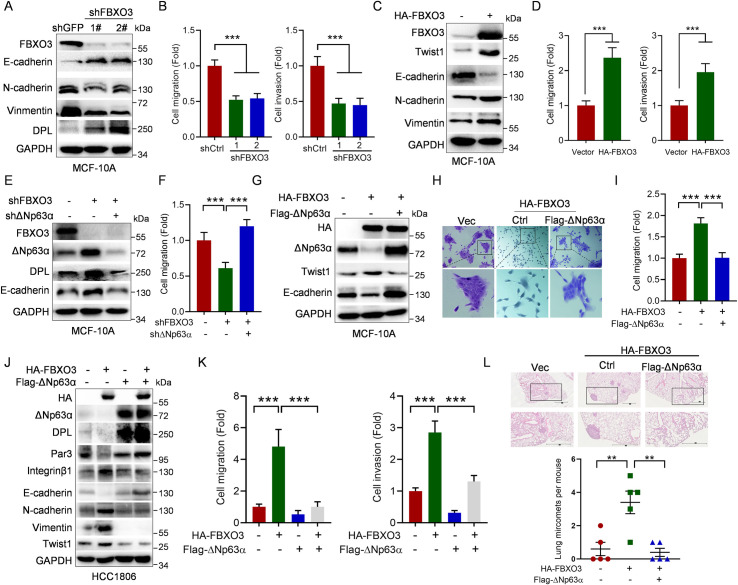
Fig 4. FBXO3 promotes tumor micrometastasis via reducing ΔNp63α protein expression.
(A, B) MCF-10A cells stably expressing 2 different shRNAs against FBXO3 (shFBXO3-1 or shFBXO3-2) or GFP (shC) were subjected to (A) western blot analyses or (B) transwell assays (migration or invasion). Data from 3 independent experiments in triplicates were presented as means ± SD. ***p < 0.001. (C, D) MCF-10A cells stably expressing HA-FBXO3 or a vector control were subjected to (C) western blot analyses or (D) transwell assays (migration or invasion). Data from 3 independent experiments in triplicates were presented as means ± SD. ***p < 0.001. (E, F) MCF-10A cells stably expressing shFBXO3-1 were infected with lentivirus expressing shΔNp63α as indicated, followed by (E) western blot analyses or (F) transwell assays. Data from 3 independent experiments in triplicates were presented as means ± SD. ***p < 0.001. (G–I) MCF-10A cells stably expressing HA-FBXO3 or a vector control were infected with lentivirus expressing Flag-ΔNp63α, followed by (G) western blot analyses, (H) representative cell morphologies, or (I) transwell assays. Data from 3 independent experiments in triplicates were presented as means ± SD. ***p < 0.001. (J, K) HCC1806 cells stably expressing HA-FBXO3 or a vector control were infected with lentivirus expressing Flag-ΔNp63α, followed by (J) western blot analyses or (K) transwell assays. Data from 3 independent experiments were presented as means ± SD. ***p < 0.001. (L) HCC1806 cells stably expressing HA-FBXO3 were infected with lentivirus expressing Flag-ΔNp63α or a vector control. The BALB/c nude mice (n = 5/group) were injected intravenously with 2 × 10⁶ HCC1806 stable cells via tail veins. Mice were euthanized by day 60 after inoculation. Paraffin embedded lungs were sectioned and stained by HE for micro nodules. The numbers of micro nodules were presented. Data were presented as means ± SEM. **p < 0.01. The underlying data for this figure can be found in S1 Data. HE, hematoxylin–eosin.

